One hour after administration to rats, noopept cannot be found in brain tissue at all. The compound that can be found — and which rises two and a half fold — is cyclo-prolylglycine, a cyclic dipeptide the brain already makes for itself. That single 1997 chromatography result reframes every subsequent mechanistic study: the question is not what noopept binds, but what its metabolite does, and whether an exogenous dose of an endogenous peptide is doing anything a receptor-level model would recognise.
Key points
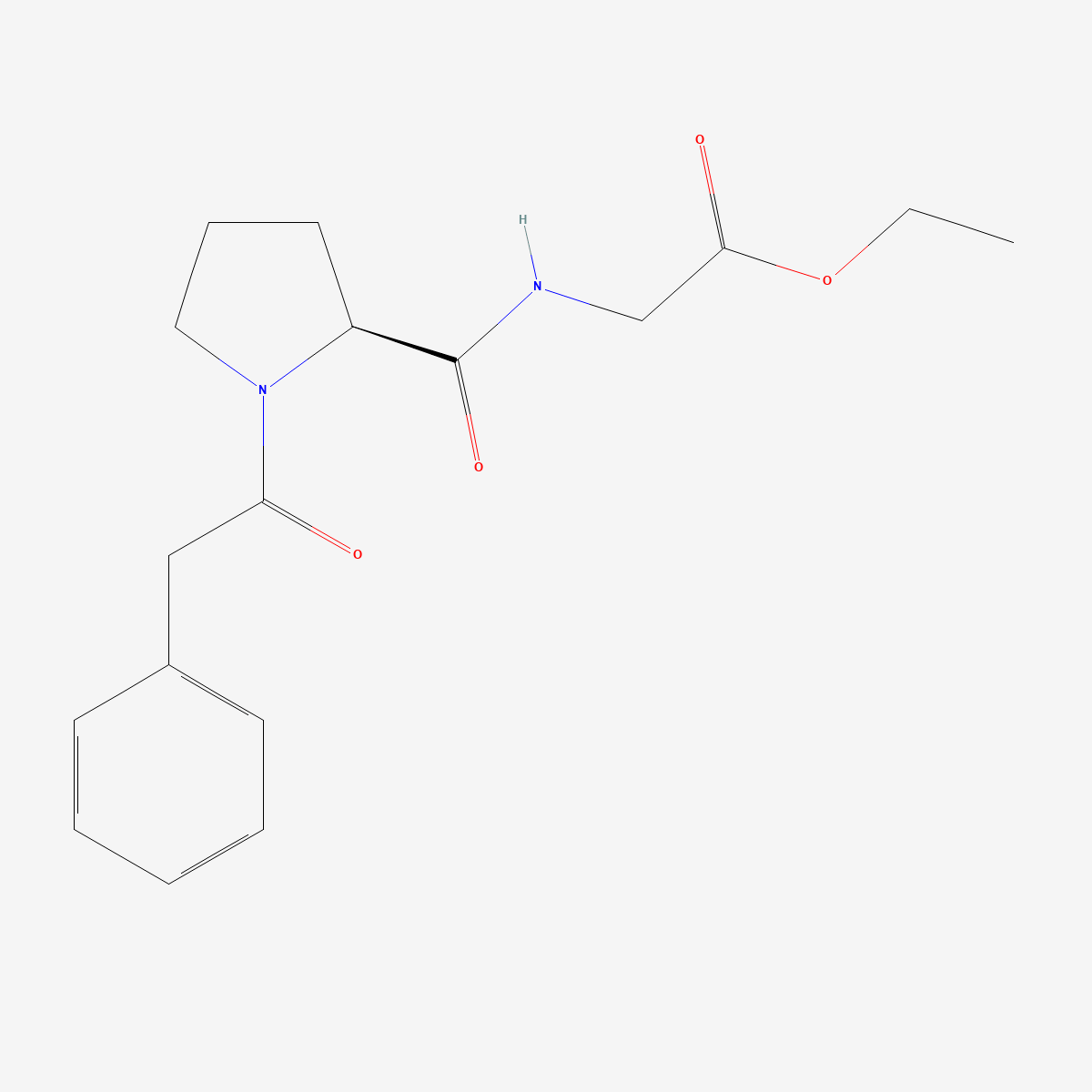
- Structure: noopept is the ethyl ester of N-phenylacetyl-L-prolylglycine (CAS 157115-85-0, C17H22N2O4, MW 318.4), also indexed as GVS-111 and omberacetam — a proline-containing dipeptide with a phenylacetyl cap and an ethyl ester tail.
- Prodrug behaviour: the parent was below the HPLC limit of detection in rat brain one hour after 5 mg/kg intraperitoneally, while cyclo-prolylglycine concentration rose 2.5-fold.[1]
- Proposed primary mechanism: selective activation of hypoxia-inducible factor 1. Of nine transcription-factor reporter constructs tested, only HIF-1 responded to 10 µM noopept; docking places the molecule in the active site of prolyl hydroxylase 2.[6]
- Neurotrophic signature: NGF and BDNF mRNA rose in rat hippocampus after acute administration, and chronic 28-day treatment potentiated rather than desensitised the effect.[3]
- Evidence base: narrow. Most primary work originates from the Zakusov Institute of Pharmacology in Moscow and its collaborators; independent replication is sparse, and the principal clinical comparison is published without an English abstract.
- Status: supplied as a reference standard for laboratory research. Not a medicine and not for human or animal consumption.
A dipeptide designed to imitate a five-membered ring
Noopept was constructed as a dipeptide analogue of piracetam — the design intent is stated explicitly in the primary literature rather than inferred.[5] The reasoning behind such a substitution is that the pyrrolidinone ring of piracetam resembles a constrained proline residue, so a proline-containing dipeptide might reproduce the relevant pharmacophore while gaining peptide-like properties. Chemically the result is not a racetam at all: there is no 2-oxopyrrolidine acetamide core, only a prolyl residue inside a linear dipeptide with an ester and an amide.[8]
That distinction matters for anyone classifying material by structure rather than by convention. Databases and vendor catalogues frequently list noopept among the racetams; the structural analysis published in 2025 makes the point that its chemical structure is quite different from other compounds of that group, piracetam included.[8] If a comparison is being drawn on scaffold similarity, the premise does not hold.
The parent compound does not reach the brain
Gudasheva and colleagues looked for GVS-111 in rat brain one hour after 5 mg/kg intraperitoneally and did not find it — not at a low concentration, but below the limit of detection of their HPLC method. Three candidate metabolites were present: phenylacetic acid, prolylglycine and cyclo-prolylglycine, all of which also occur in control animals. Only cyclo-prolylglycine increased, by a factor of 2.5. Formation of cyclo-prolylglycine from the parent was then reproduced in vitro in the presence of plasma and brain enzymes.[1]
The authors concluded that the compound is a prodrug converting in the body to a cyclopeptide identical to the endogenous one. Pharmacokinetic work has since tracked the parent and its metabolite together.[7] The practical consequence for experimental design is substantial: an in vitro preparation lacking the relevant esterase and cyclisation activity may be testing a molecule that never forms in vivo, and any binding screen run on the parent compound is answering a question about the prodrug rather than about the active species.
A transcriptional hypothesis, tested against eight alternatives
The most direct attempt to identify a primary mechanism is Vakhitova and colleagues’ reporter screen in HEK293 cells transiently transfected with luciferase constructs for CREB, NFAT, NF-κB, p53, STAT1, GAS, VDR, HSF1 and HIF-1. Noopept at 10 µM increased DNA-binding activity of HIF-1 only, leaving the other eight unchanged, and produced a further increase under cobalt chloride-induced HIF-1 stabilisation in a concentration-dependent manner. Piracetam at 1 mM — a hundredfold higher concentration — affected none of the factors tested.[6]
Molecular docking in the same study placed the L-isomer of noopept, and the L-isomer of its N-phenylacetylprolyl metabolite, in the active site of prolyl hydroxylase 2, while the pharmacologically inactive D-isomer did not dock. That is an internally consistent story: inhibit the prolyl hydroxylase that marks HIF-1α for degradation, and HIF-1 accumulates. The stereochemical dissociation is the part worth noting, because it is the kind of control that distinguishes a mechanism from a correlation. The authors proposed the HIF-positive effect as the primary mechanism underlying the wider spectrum of reported effects.
Neurotrophins, and an effect that grows rather than fades
Ostrovskaya and colleagues measured NGF and BDNF mRNA in rat cerebral cortex and hippocampus by Northern blot after single and 28-day administration. The pattern was regionally split: in cortex, expression of both neurotrophins fell below control after a single dose, with a slight increase in BDNF after chronic treatment; in hippocampus, both rose after acute administration, and chronic treatment potentiated rather than attenuated the effect.[3]
Absence of tolerance over 28 days is an unusual finding for a compound acting on a signalling pathway, and it fits a transcriptional mechanism better than a receptor-occupancy one. It is also a constraint on assay design: an experiment sampling at a single early timepoint in cortex would report the opposite sign to one sampling hippocampus, which is a plausible source of irreproducibility across laboratories using different dissections.
Cell-model neuroprotection, including work from outside the originating network
The most useful independent contribution comes from a collaboration involving the University of Connecticut Health Center. Pelsman and colleagues tested GVS-111 in normal human cortical neurons treated with 50 µM hydrogen peroxide, a treatment that degenerated more than 60% of neurons in culture, and in cortical neurons from Down’s syndrome tissue. The compound increased neuronal survival with dose-dependent activity from 10 nM to 100 µM and an IC50 of 1.21 ± 0.07 µM, inhibited accumulation of intracellular free radicals and lipid peroxidation, and outperformed piracetam as well as the antioxidants vitamin E, propyl gallate and s-PBN in the same assay.[2]
Later work in the amyloid-β(25–35) PC12 model reported reduced apoptosis, lower intracellular reactive oxygen species and calcium, restored mitochondrial membrane potential, and attenuated tau phosphorylation at Ser396, with the compound applied at 10 µM for 72 hours before a 5 µM peptide challenge.[5] A separate patch-clamp study identified a cholinergic route: 5 µM noopept increased action-potential firing in GABAergic interneurons of hippocampal stratum radiatum, and that increase was almost completely abolished by the α7 nicotinic antagonists α-bungarotoxin at 6 nM and methyllycaconitine at 20 nM.[4]
How narrow the evidence base actually is
Three limitations belong in any honest summary. First, authorship is concentrated: Gudasheva, Ostrovskaya and Seredenin appear on the synthesis paper, the metabolism paper, the neurotrophin paper, the cell-model papers and the electrophysiology paper. That is normal for a compound developed inside one institute, but it means the findings are not independent observations in the statistical sense.
Second, the clinical literature is thin and largely inaccessible. The principal comparison against piracetam in patients with mild cognitive disorders of vascular and traumatic origin is indexed in PubMed without an English abstract,[9] which means its design, sample size and effect size cannot be verified from the index record. We are not in a position to state what it found, and we do not.
Third, the HIF-1 mechanism is proposed rather than established. It rests principally on one reporter-construct study in a heterologous cell line plus docking, and no independent laboratory appears to have reproduced the selectivity across those nine transcription factors. It is the best-supported mechanistic hypothesis available for this molecule, which is a different statement from saying it is the mechanism.
Practical notes for laboratory handling
The ethyl ester is the obvious liability. Esterases in serum-containing media will cleave it, so a cell-culture experiment intending to test the parent compound should account for hydrolysis over the incubation period rather than assume the nominal concentration persists — particularly in the 72-hour pre-treatment designs used in the amyloid work. Aqueous stock solutions should be prepared fresh.
Solid-state characterisation is now available: the 2025 analysis combined TGA/DSC, solution-state 1H and 13C NMR, 13C CP/MAS NMR, scanning electron microscopy, single-crystal and powder X-ray diffraction with periodic DFT computation, and included a polymorphism screen.[8] That is the reference point for identity confirmation by powder diffraction, and it is worth using, because until that paper appeared very little was published on the physical form of this material.
Frequently asked questions
Is noopept a racetam?
By convention it is often grouped with them, because it was deliberately designed as a dipeptide analogue of piracetam.[5] By structure it is not: it lacks the 2-oxopyrrolidine acetamide core, and the structural analysis published in 2025 states plainly that its chemistry differs considerably from the rest of that group.[8] If the classification is being used to predict behaviour in an assay, the structural reading is the safer one.
Why is noopept reported active at so much lower concentrations than piracetam?
The measured contrast is real in at least one head-to-head setting: 10 µM noopept moved HIF-1 reporter activity where 1 mM piracetam moved nothing among nine transcription factors tested.[6] A similar gap appeared in human cortical neurons, where noopept showed significantly higher neuroprotection than piracetam against peroxide-induced damage.[2] Whether the gap reflects a different mechanism rather than a more efficient one is not resolved by those experiments.
Has noopept been studied outside Russia?
Some. The human cortical neuron work involved a United States laboratory,[2] and the physicochemical characterisation came from groups in Poland and Greece.[8] The mechanistic core of the literature, however, remains concentrated in one Moscow institute and its partner institute in Ufa, and no large independent replication of the behavioural or clinical findings has been published.
What purity is appropriate for a reference standard?
For peptide-chemistry work the relevant impurities are the free acid from ester hydrolysis and the D-isomer, which the docking study indicates is pharmacologically inactive. Material characterised at ≥99% with a stated assay method, and ideally with a chiral purity figure, is the practical minimum. Our noopept reference material (CAS 157115-85-0), assayed at ≥99% purity, ships with identity documentation available on request.
Related compounds in our catalogue
Laboratories benchmarking this dipeptide against the scaffold it was modelled on generally work alongside aniracetam reference standard (CAS 72432-10-1), an anisoyl-substituted pyrrolidinone, and pramiracetam reference material (CAS 68497-62-1), a piracetam derivative carrying a diisopropylaminoethyl amide — both retaining the 2-oxopyrrolidine core that noopept does not have.
References
- Gudasheva TA, Boyko SS, Ostrovskaya RU, Voronina TA, Akparov VK, Trofimov SS, Rozantsev GG, Skoldinov AP, Zherdev VP, Seredenin SB. The major metabolite of dipeptide piracetam analogue GVS-111 in rat brain and its similarity to endogenous neuropeptide cyclo-L-prolylglycine. European Journal of Drug Metabolism and Pharmacokinetics. 1997;22(3):245–252. doi:10.1007/BF03189814 · PMID: 9358206
- Pelsman A, Hoyo-Vadillo C, Gudasheva TA, Seredenin SB, Ostrovskaya RU, Busciglio J. GVS-111 prevents oxidative damage and apoptosis in normal and Down’s syndrome human cortical neurons. International Journal of Developmental Neuroscience. 2003;21(3):117–124. doi:10.1016/s0736-5748(03)00031-5 · PMID: 12711349
- Ostrovskaya RU, Gudasheva TA, Zaplina AP, Vahitova JV, Salimgareeva MH, Jamidanov RS, Seredenin SB. Noopept stimulates the expression of NGF and BDNF in rat hippocampus. Bulletin of Experimental Biology and Medicine. 2008;146(3):334–337. doi:10.1007/s10517-008-0297-x · PMID: 19240853
- Kondratenko RV, Povarov IS, Kolbaev SN, Derevyagin VI, Ostrovskaya RU, Gudasheva TA, Sharonova IN, Skrebitsky VG. Effect of nootropic dipeptide noopept on CA1 pyramidal neurons involves α7AChRs on interneurons in hippocampal slices from rat. Neuroscience Letters. 2022;790:136898. doi:10.1016/j.neulet.2022.136898 · PMID: 36195298
- Ostrovskaya RU, Vakhitova YV, Kuzmina USh, Salimgareeva MKh, Zainullina LF, Gudasheva TA, Vakhitov VA, Seredenin SB. Neuroprotective effect of novel cognitive enhancer noopept on AD-related cellular model involves the attenuation of apoptosis and tau hyperphosphorylation. Journal of Biomedical Science. 2014;21(1):74. doi:10.1186/s12929-014-0074-2 · PMID: 25096780
- Vakhitova YV, Sadovnikov SV, Borisevich SS, Ostrovskaya RU, Gudasheva TA, Seredenin SB. Molecular mechanism underlying the action of substituted Pro-Gly dipeptide Noopept. Acta Naturae. 2016;8(1):82–89. PMID: 27099787
- Boyko SS, Zherdev VP, Shevchenko RV. Pharmacokinetics of noopept and its active metabolite cycloprolyl glycine in rats. Biomeditsinskaia Khimiia. 2018;64(5):455–458. doi:10.18097/PBMC20186405455 · PMID: 30378564
- Araj SK, Szeleszczuk Ł, Gubica T, Zielińska-Pisklak M, Bethanis K, Christoforides E, Dudek MK, Pisklak DM. Physicochemical and structural analysis of N-phenylacetyl-L-prolylglycine ethyl ester (Noopept) — an active pharmaceutical ingredient with nootropic activity. Journal of Pharmaceutical and Biomedical Analysis. 2025;252:116474. doi:10.1016/j.jpba.2024.116474 · PMID: 39298839
- Neznamov GG, Teleshova ES. Comparative studies of Noopept and piracetam in the treatment of patients with mild cognitive disorders in organic brain diseases of vascular and traumatic origin. Neuroscience and Behavioral Physiology. 2009;39(3):311–321. doi:10.1007/s11055-009-9128-4 · PMID: 19234797
Research use only. The compounds discussed are supplied as reference standards for laboratory research. They are not medicines, not dietary supplements, and are not for human or animal consumption. Nothing in this article is medical advice or a recommendation for use in humans. Any quantities mentioned are those reported in the published studies cited above and appear solely to describe that research accurately.