L-DOPA does not bind a dopamine receptor. It is a substrate, not a ligand, and on its own it does essentially nothing — which means its entire pharmacology is decided by a question of location: where the decarboxylase that converts it happens to sit. Gut bacteria, intestinal wall, peripheral tissue and brain all carry that enzymatic capacity, and the fraction converted at each site determines everything from bioavailability to the involuntary movements that limit its long-term use. Sixty years after the first clinical successes, two of the most informative results on this molecule were published in the last decade.
Key points
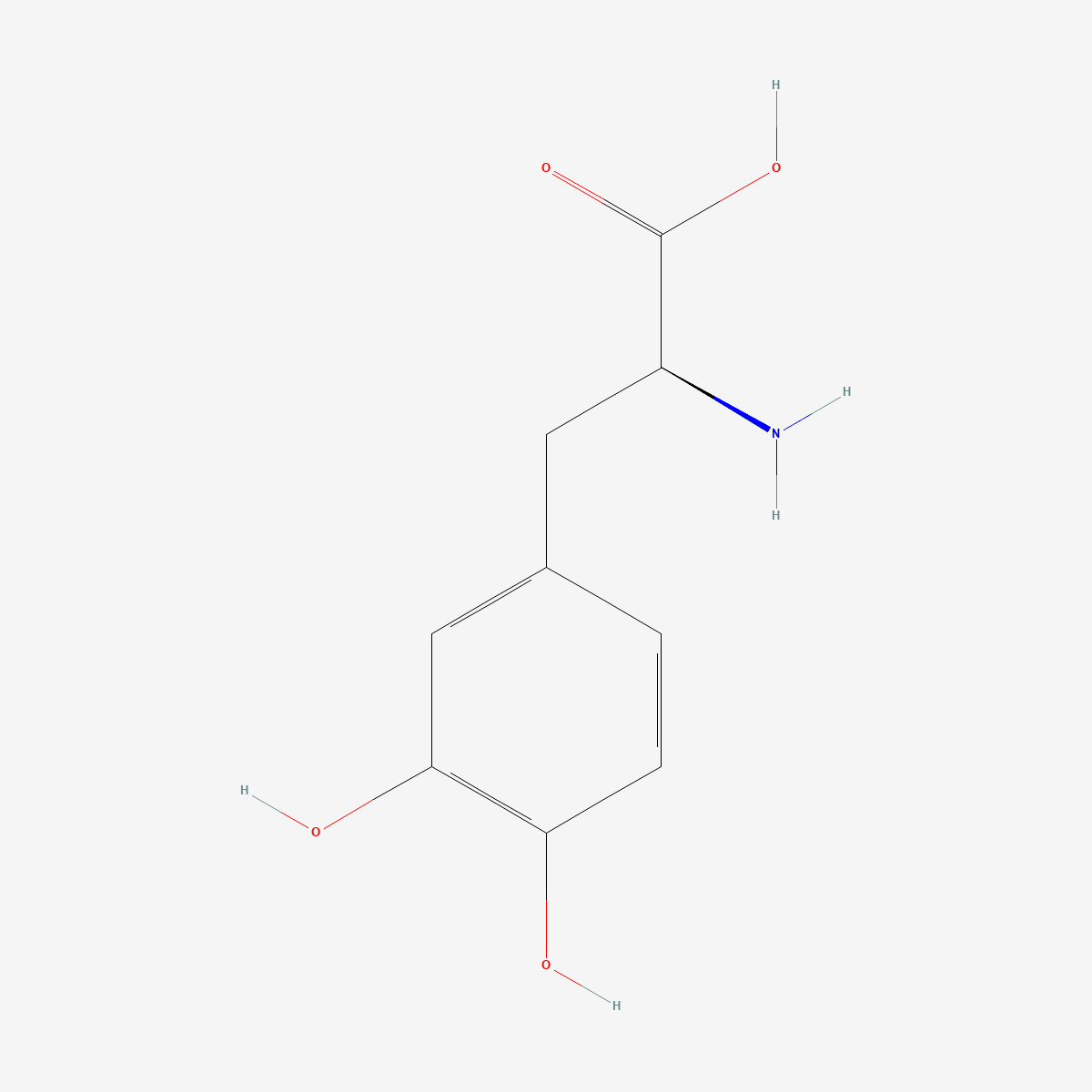
- Structure: L-DOPA is (S)-2-amino-3-(3,4-dihydroxyphenyl)propanoic acid (CAS 59-92-7, C9H11NO4, MW 197.19) — a catechol fused to an α-amino acid backbone, which is exactly why it crosses membranes that dopamine cannot.
- Not directly active: the molecule requires decarboxylation by aromatic L-amino acid decarboxylase (AADC) to produce dopamine; it does not mimic a dopaminergic agonist.[6]
- Transport is saturable and competitive: absorption in the proximal small bowel uses a facilitated transport system shared with other large neutral amino acids, and the same carriers move the molecule from plasma into brain.[2]
- A bacterial decarboxylase competes for the substrate: a pyridoxal phosphate-dependent tyrosine decarboxylase from Enterococcus faecalis converts L-DOPA to dopamine in the gut, and a drug targeting the host enzyme does not prevent it.[3]
- Decarboxylase capacity is rate-limiting: putaminal AADC gene therapy increased the motor response to a fixed intravenous levodopa infusion by 168% in a controlled substudy — and increased dyskinesia scores by 208%.[5]
- Status: supplied as a reference standard for laboratory research. Not a medicine and not for human or animal consumption.
A catechol amino acid, and why that combination matters
Dopamine and L-DOPA differ by one carboxyl group. That group is the whole story. Dopamine is a catecholamine with no amino acid character and is not a substrate for the carriers that move amino acids across the blood-brain barrier; L-DOPA retains the α-amino acid motif and is carried by the large neutral amino acid transport system that handles phenylalanine, tyrosine, leucine and the rest.[2] Adding a carboxyl to a neurotransmitter to make it transportable, then removing it enzymatically at the destination, is prodrug design that predates the concept being named.
The competitive consequence is often underappreciated. Because the carrier is shared and saturable, the other large neutral amino acids in circulation are competitors, not bystanders. Pharmacokinetic reviews accordingly list standardisation of dosing with respect to meal times and control of dietary protein among the principal strategies for making intestinal absorption and brain delivery reproducible.[2] For an in vivo experiment, the amino acid composition of the diet is an experimental variable in the same sense that the dose is.
The historical argument was about dose, not about mechanism
The mechanistic groundwork was laid quickly and the clinical translation took a decade longer, for reasons worth understanding. A 1957 experiment showed that L-DOPA could reverse reserpine-induced immobility in animals; the same laboratory then showed that reserpine depleted brain dopamine and that L-DOPA restored it, and that dopamine was most concentrated in the striatum. A 1960 postmortem study demonstrated marked striatal dopamine depletion in Parkinson’s disease, and the first patient injections followed.[1]
Those early trials produced limited and short-lived responses, and several groups failed to find any benefit at all. The resolution came from dose. Cotzias reported in 1967 that d,l-dopa reversed symptoms dramatically, but only at extremely high dosages and with considerable haematologic adverse effects; switching to the L-enantiomer gave the same benefit at half the dosage and without the haematologic problems, and an independent double-blind trial then confirmed it.[1]
That sequence is a clean illustration of why the location question dominates. Almost all of an oral dose is decarboxylated before it reaches the brain, so the early low-dose failures were not testing the hypothesis they appeared to be testing. The modern answer — co-administering an inhibitor of extracerebral AADC, which cannot itself cross into the brain — achieves the same end without the dose escalation.[2]
A second decarboxylase nobody had accounted for
The presystemic loss turned out to have a component that no host-directed inhibitor addresses. Maini Rekdal and colleagues described an interspecies pathway in the human gut microbiota: conversion of L-DOPA to dopamine by a pyridoxal phosphate-dependent tyrosine decarboxylase from Enterococcus faecalis, followed by transformation of that dopamine to m-tyramine by a molybdenum-dependent dehydroxylase from Eggerthella lenta. These enzymes predicted drug metabolism across complex human gut microbiota samples. Critically, a drug targeting host AADC did not prevent the bacterial decarboxylation; the authors then identified a separate compound that inhibited the bacterial activity in patient-derived microbiota and increased bioavailability in mice.[3]
Beckers, Bloem and Verbeek set this alongside a second route to the same outcome. They describe two mechanisms of peripheral resistance: impaired bowel motility leading to bacterial overgrowth and excessive bacterial tyrosine decarboxylase, and systemic induction of host AADC causing premature conversion. Both reduce the fraction arriving in brain, and both are invisible to an analysis that treats the administered dose as the exposure.[4]
For anyone working with this compound in an animal model, the implication is uncomfortable but concrete: two cohorts with different microbiota can receive identical doses and experience different exposures, and the difference will look like biological variability rather than what it is.
Direct evidence that decarboxylase capacity sets the ceiling
The cleanest test of the central claim is a surgical one. In the PD-1101 study, an adeno-associated virus serotype-2 vector carrying the AADC gene was administered into the putamen of patients with advanced Parkinson’s disease — that is, the decarboxylase capacity was increased directly while everything upstream was left alone. Thirteen of fifteen patients took part in a substudy in which intravenous levodopa was infused at threshold (0.6 mg/kg/h) and suprathreshold (1.2 mg/kg/h) rates, under randomised and double-blinded infusion order, before and approximately six months after vector administration.[5]
Motor score area-under-curve responses increased by 168% and 67% at the two infusion rates; finger-tapping speeds improved by 162% and 113%. The same intervention increased dyskinesia scores by 208% and 72%. That symmetry is the point rather than a caveat: if adding decarboxylase amplifies both the therapeutic response and the principal dose-limiting adverse effect to the same fixed substrate load, then the enzyme is genuinely the bottleneck, and the two outcomes are two readings of one mechanism.
Once inside the brain, the wrong cells may be doing the releasing
Decarboxylation in the brain does not mean decarboxylation in the right place. AADC is not confined to dopaminergic terminals, and as those terminals degenerate their share of total capacity falls. Chagraoui and colleagues review the consequence: L-DOPA-derived dopamine is mainly released by serotonergic neurons as a false neurotransmitter, serotonergic neurons are implicated in levodopa-induced dyskinesia, and the pattern and magnitude of extracellular dopamine achieved by this route differ from physiological dopaminergic transmission. They further note that trace amines and other derivatives formed from the newly made dopamine, or from the parent molecule directly, can accumulate in monoaminergic terminals and impair exocytotic release of the native monoamines.[6]
A 2025 review frames the same phenomenon in terms of buffering. A healthy dopaminergic terminal stores and releases dopamine on demand, smoothing the relationship between plasma concentration and synaptic concentration; as those terminals are lost, that buffering capacity goes with them, striatal dopamine concentrations begin to track the timing of administration directly, and reliance on serotonergic false-neurotransmitter release increases.[7] The short plasma half-life, which is tolerable while buffering is intact, becomes the dominant variable once it is not.
This is why mechanistic descriptions that stop at “L-DOPA replaces missing dopamine” are misleading rather than merely simplified. The molecule supplies substrate; which cells decarboxylate it, store it and release it is determined by the state of the tissue, and that state changes over the course of the disease being modelled.
Practical notes for laboratory handling
The catechol is the vulnerability. Catechols autoxidise readily in neutral and alkaline aqueous solution, accelerated by dissolved oxygen, light and trace transition metals, with quinone and melanin-type oligomers as the visible endpoint — solutions darken from colourless through pink to brown. A discoloured stock is a chemically different reagent, not merely an aged one, and in an oxidative-stress assay the degradation products may be the more active species. Prepare solutions fresh in degassed acidified buffer, protect from light, and treat colour as a rejection criterion rather than a cosmetic observation. The free amino acid is sparingly soluble in water at neutral pH, which is why dissolution is usually performed under acidic conditions before adjustment.
Frequently asked questions
Why is L-DOPA used rather than dopamine itself?
Because dopamine is not carried across the blood-brain barrier, whereas L-DOPA retains the α-amino acid structure recognised by the large neutral amino acid transport system, and the same carriers operate in the intestinal mucosa and at the barrier.[2] The carboxyl group that makes transport possible is then removed enzymatically at the destination.
Why does inhibiting decarboxylase outside the brain increase the effect inside it?
Because the two compartments compete for the same substrate. Extensive presystemic metabolism consumes most of an oral dose before it reaches the central compartment; inhibitors that cannot cross into the brain suppress that peripheral conversion without suppressing the conversion that is wanted.[2] The strategy is incomplete, however, because the gut bacterial decarboxylase is a different enzyme and is not blocked by host-directed inhibitors.[3]
Do gut bacteria really change how much L-DOPA reaches the brain?
The enzymology is established: a specific Enterococcus faecalis tyrosine decarboxylase and a specific Eggerthella lenta dehydroxylase account for the conversion, they predict metabolism across complex human microbiota samples, and selectively inhibiting the bacterial enzyme increased bioavailability in mice.[3] Whether this explains a given individual’s variable response is a separate and less settled question, addressed as one of two candidate mechanisms in reviews of peripheral resistance.[4]
What purity is appropriate for a reference standard?
Enantiomeric purity matters as much as chemical purity here: the historical dose comparison between the racemate and the resolved L-form was decided on toxicity, not on potency alone.[1] Oxidation products, being coloured and redox-active, are the other impurity class to specify. Material characterised at ≥99% with a stated assay method and a chiral purity figure is the practical minimum. Our L-DOPA reference standard (CAS 59-92-7), assayed at ≥99% purity, ships with identity documentation available on request; enzyme activity in AADC-deficient patients and heterozygous carriers has also been characterised in the clinical literature, which gives a reference range for assay work.[8]
Related compounds in our catalogue
Precursor loading as a pharmacological strategy is not confined to dopamine, and laboratories comparing the approach across transmitter systems commonly hold alpha-GPC reference material (CAS 28319-77-9), a choline precursor studied on the same logic of supplying substrate rather than engaging a receptor. For the opposite end of dopamine handling — clearance rather than synthesis — modafinil reference material (CAS 68693-11-8) acts at the dopamine transporter that removes the neurotransmitter this molecule supplies.
References
- Lees AJ, Tolosa E, Olanow CW. Four pioneers of L-dopa treatment: Arvid Carlsson, Oleh Hornykiewicz, George Cotzias, and Melvin Yahr. Movement Disorders. 2015;30(1):19–36. doi:10.1002/mds.26120 · PMID: 25488030
- Contin M, Martinelli P. Pharmacokinetics of levodopa. Journal of Neurology. 2010;257(Suppl 2):S253–S261. doi:10.1007/s00415-010-5728-8 · PMID: 21080186
- Maini Rekdal V, Bess EN, Bisanz JE, Turnbaugh PJ, Balskus EP. Discovery and inhibition of an interspecies gut bacterial pathway for Levodopa metabolism. Science. 2019;364(6445):eaau6323. doi:10.1126/science.aau6323 · PMID: 31196984
- Beckers M, Bloem BR, Verbeek MM. Mechanisms of peripheral levodopa resistance in Parkinson’s disease. npj Parkinson’s Disease. 2022;8(1):56. doi:10.1038/s41531-022-00321-y · PMID: 35546556
- Nutt JG, Curtze C, Hiller A, Anderson S, Larson PS, Van Laar AD, Richardson RM, Thompson ME, Sedkov A, Leinonen M, Ravina B, Bankiewicz KS, Christine CW. Aromatic L-amino acid decarboxylase gene therapy enhances levodopa response in Parkinson’s disease. Movement Disorders. 2020;35(5):851–858. doi:10.1002/mds.27993 · PMID: 32149427
- Chagraoui A, Boulain M, Juvin L, Anouar Y, Barrière G, Deurwaerdère P. L-DOPA in Parkinson’s disease: looking at the “false” neurotransmitters and their meaning. International Journal of Molecular Sciences. 2019;21(1):294. doi:10.3390/ijms21010294 · PMID: 31906250
- Riederer P, Strobel S, Nagatsu T, Watanabe H, Chen X, Löschmann PA, Sian-Hulsmann J, Jost WH, Müller T, Dijkstra JM, Monoranu CM. Levodopa treatment: impacts and mechanisms throughout Parkinson’s disease progression. Journal of Neural Transmission. 2025;132(6):743–779. doi:10.1007/s00702-025-02893-4 · PMID: 40214767
- Verbeek MM, Geurtz PB, Willemsen MA, Wevers RA. Aromatic L-amino acid decarboxylase enzyme activity in deficient patients and heterozygotes. Molecular Genetics and Metabolism. 2007;90(4):363–369. doi:10.1016/j.ymgme.2006.12.001 · PMID: 17240182
Research use only. The compounds discussed are supplied as reference standards for laboratory research. They are not medicines, not dietary supplements, and are not for human or animal consumption. Nothing in this article is medical advice or a recommendation for use in humans. Any quantities mentioned are those reported in the published studies cited above and appear solely to describe that research accurately.