In rats given a single oral dose of radiolabelled apigenin, radioactivity does not appear in blood until 24 hours have passed, and the terminal elimination half-time is 91.8 hours. In humans, the aglycone is absorbed so poorly that urinary metabolites account for 0.5% of what was swallowed. Those two statements look contradictory — almost nothing gets in, yet what gets in stays for days — and reconciling them is the whole of this molecule’s pharmacokinetics. The resolution involves a gut wall that conjugates faster than the liver, a microbial population that decides how much aglycone is ever liberated, and a set of circulating species that are not apigenin.
Key points
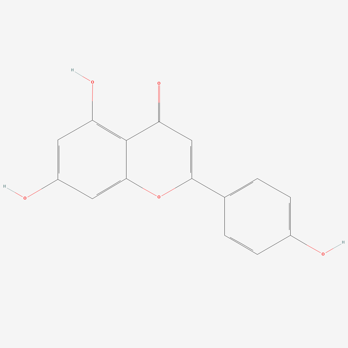
- Identity: apigenin is 5,7,4′-trihydroxyflavone (CAS 520-36-5, C15H10O5, MW 270.24), the aglycone; its glycosides apiin and apigenin-7-O-glucoside are different chemical entities with different absorption behaviour.
- Human absorption of the aglycone: apigenin per se was poorly absorbed, with metabolites equivalent to 0.5% of intake excreted in urine over 0–24 h.[3] Two independent parsley studies put urinary recovery of free apigenin at 0.22 ± 0.16%[4] and 0.58 ± 0.16%[5] of intake.
- What circulates: apigenin-4′-glucuronide, apigenin-7-glucuronide and apigenin-7-sulfate — conjugates, not free aglycone.[3]
- Rat disposition: after a single oral dose, 51.0% of radioactivity in urine, 12.0% in faeces and 24.8% still in the rest of the body at 10 days; elimination half-time 91.8 h, distribution volume 259 mL, plasma clearance 1.95 mL/h.[1]
- The matrix dominates the number: chamomile tea gave a 2 h plasma peak and urinary metabolite recovery equal to 34% of intake; a parsley drink gave a 4 h peak and 11.2%; the aglycone itself gave 0.5%.[3]
- Status: supplied as a reference standard for laboratory research. Not a medicine, not a dietary supplement, and not for human or animal consumption.
The mass balance, which is more informative than any single parameter
The cleanest description of apigenin disposition comes from a radiolabel study in female and male rats. After a single oral administration, recovery over ten days was distributed as 51.0% of radioactivity in urine, 12.0% in faeces, 1.2% in blood, 0.4% in kidneys, 9.4% in intestine, 1.2% in liver and 24.8% in the rest of the body. Radioactivity appeared in blood only 24 hours after dosing. Blood kinetics gave an elimination half-time of 91.8 h, a distribution volume of 259 mL and a plasma clearance of 1.95 mL/h, and the authors concluded that absorption and elimination are both slow, raising the possibility of accumulation.[1]
Read the numbers rather than the summary and two things stand out. First, roughly a quarter of the administered radioactivity was still in the body at ten days — that figure is what drives the long half-time, and it describes label rather than necessarily intact apigenin. Second, the 24-hour delay before blood radioactivity appears is not the behaviour of a small lipophilic molecule crossing the duodenal wall. It is the behaviour of a compound whose absorption happens distally, after something in the gut has acted on it.
The same study found a sex difference in which conjugate dominates. Immature male and female rats, like mature females, excreted a higher percentage of the mono-glucuronoconjugate than of the mono-sulfoconjugate (10.0–31.6% versus 2.0–3.6%); mature males excreted the same compounds in the inverse ratio, 4.9% and 13.9%.[1] The conjugation profile is therefore partly a function of the animals chosen, not a fixed property of the compound.
Phase I produces another flavone that is sold separately
Apigenin’s phase I metabolism has a feature that most flavone metabolism does not: its principal oxidative product is itself a well-known compound. In rat liver subcellular fractions, phase I metabolism gave three monohydroxylated derivatives — luteolin as the major metabolite (Km 22.5 ± 1.5 µM, Vmax 5.605 ± 0.090 nmol/min/mg protein), together with scutellarein and iso-scutellarein. Inhibitor and inducer experiments implicated CYP1A1, CYP2B and CYP2E1. In parallel, phase II metabolism in vitro produced three monoglucuronoconjugates and one monosulfoconjugate of apigenin, while luteolin itself gave four monoglucuronoconjugates, two sulfoconjugates and one methylconjugate identified as diosmetin.[2]
The ex vivo arm of that study is the part with practical consequences. When apigenin was perfused through an isolated rat liver, none of the phase I metabolites could be recovered; what emerged were two monoglucuronoconjugates and one sulfoconjugate already seen in vitro, plus a diglucuronoconjugate and a glucuronosulfoconjugate.[2] In an intact organ, conjugation outruns oxidation so completely that the hydroxylation products never accumulate.
This matters for identity work as much as for kinetics. Luteolin differs from apigenin by a single hydroxyl, co-elutes under short gradients, and is both a plausible starting-material impurity and a documented metabolite. Only the provenance of the sample distinguishes those two explanations of the same peak.
Which enzymes, and where
The conjugating enzymology has been resolved to the level of individual positions on the ring. Human liver and intestinal microsomes both form two glucuronides from apigenin, AP-7G and AP-4’G. Total glucuronidation activity was higher in liver than in intestinal microsomes, but the regioselectivity inverted between the two tissues: intrinsic clearance ran AP-7G > AP-4’G in liver microsomes and AP-7G < AP-4’G in intestinal microsomes.[8] Those same two glucuronides are among the species identified in human plasma and urine,[3] so the ratio between them carries information about which tissue did the conjugating.
Sulfation has been mapped across the human cytosolic sulfotransferase family. Of the thirteen human SULTs examined, four — 1A1, 1A2, 1A3 and 1C4 — displayed sulfating activity towards apigenin, and among four human organ specimens, liver and intestine cytosols showed much higher activity than lung and kidney.[9] Liver and gut again, rather than a peripheral tissue.
Enteric recycling: the gut wall outperforms the liver
The reason a compound with reasonable membrane permeability ends up with single-digit-percent systemic availability is that it is conjugated and re-excreted before it ever leaves the intestine. A four-site perfused rat intestinal model quantified this directly. Approximately 28% of perfused aglycones were absorbed, roughly 450 nmol per 30 min. Maximal intestinal conjugate excretion per 30 min was 150 nmol for apigenin against 61 nmol for genistein, while maximal biliary conjugate excretion was 30 nmol for apigenin against 50 nmol for genistein. Microsomes from Caco-2 cells, rat intestine and rat liver always glucuronidated apigenin faster than genistein, with rat jejunal microsomes fastest of the intestinal regions and often outpacing liver microsomes. The authors concluded that intestinal disposition may matter more than hepatic disposition in flavonoid first-pass metabolism, and that enteric recycling in conjunction with enterohepatic recycling explains the poor systemic bioavailability of this class.[7]
Note the direction of that comparison: for apigenin, more conjugate goes into the gut lumen and less into bile. The molecule is pushed back where it came from rather than routed through the liver, which is why reasoning from hepatic clearance under-predicts its first-pass loss.
The microbiota decide how much aglycone ever exists
Almost no dietary apigenin arrives as the aglycone. It arrives as apiin, apigenin-7-O-glucoside or a related glycoside, and something has to remove the sugar. A comparison of germ-free and human microbiota-associated rats given apigenin-7-glucoside established how much of that hydrolysis is microbial. In vitro, human faecal suspensions converted the glucoside to apigenin, naringenin and 3-(4-hydroxyphenyl)propionic acid. In germ-free rats, apigenin, luteolin and their conjugates appeared in urine and faeces. In the microbiota-associated animals, naringenin, eriodictyol, phloretin and a set of hydroxyphenylpropionic and hydroxycinnamic acids were formed in addition, in free and conjugated forms.[6]
The total quantity recovered barely moved — 11% of dose in germ-free animals and 13% in colonised ones over 48 h — but the route and the identity of what was recovered changed completely. Germ-free animals excreted mainly into faeces, dominated by apigenin and its conjugates; colonised animals excreted predominantly into urine, with 3-(4-hydroxyphenyl)propionic acid as the main metabolite. Deglycosylation activity was demonstrated in cytosolic extracts of Eubacterium ramulus and Bacteroides distasonis and, separately, in the microsomal fraction of rat small intestinal mucosa.[6]
That last detail matters: deglycosylation is not exclusively microbial, since host mucosal enzymes can do it too, which is presumably why germ-free animals absorbed anything at all. The microbiota shift the balance, the ring-fission chemistry and the excretion route rather than switching absorption on and off.
Human exposure, and why the food matrix dominates the number
Three human datasets exist, and the places where they disagree are instructive rather than contradictory. The most complete is a controlled study in healthy male adults which identified apigenin-4′-glucuronide, apigenin-7-glucuronide and apigenin-7-sulfate as the in vivo metabolites. Apigenin per se was poorly absorbed, with metabolites equivalent to 0.5% of intake excreted in urine 0–24 h. A parsley drink containing apigenin-7-O-(2″-O-apiosyl)glucoside gave a plasma Cmax for the 4′-glucuronide at 4 h, which the authors read as absorption in the lower gastrointestinal tract, with urinary excretion of the three metabolites corresponding to 11.2% of intake; dried parsley leaves with yogurt extended that Cmax to 6 h. Chamomile tea containing apigenin-7-O-glucoside gave a 2 h Cmax, consistent with absorption in the upper gastrointestinal tract, and urinary excretion equivalent to 34% of intake.[3]
A separate bolus study in eleven healthy subjects aged 23–41, given 2 g of blanched parsley per kilogram body weight after an apigenin- and luteolin-free diet — corresponding to 65.8 ± 15.5 µmol apigenin — measured a maximum plasma apigenin concentration of 127 ± 81 nmol/L reached after 7.2 ± 1.3 h, with wide inter-individual variation. Plasma concentrations fell below the 2.3 nmol/L detection limit within 28 h, and average 24-hour urinary apigenin of 144 ± 110 nmol corresponded to 0.22 ± 0.16% of the ingested dose.[4] A randomised crossover in seven men and seven women on a strictly controlled low-flavone diet put the excreted fraction at 0.58 ± 0.16% of intake during parsley intervention, with urinary apigenin of 1.59–409.09 µg/MJ per 24 h against 0–112.27 µg/MJ on the basic diet.[5]
The spread between 0.22%, 0.58%, 11.2% and 34% is not measurement noise. It tracks the chemical form ingested, the site of absorption implied by the time to peak, and whether free aglycone or summed conjugates were quantified: the sub-percent figures come from measuring free apigenin in urine, the double-digit figures from summing conjugates after a glycoside. Any single-number statement of apigenin bioavailability is under-specified unless it says which of those it means.
What this means for experimental design
Most in vitro pharmacology for this flavone is run at free aglycone concentrations of roughly 1–10 µM. Human plasma after a substantial parsley bolus peaked at 127 nmol/L[4] — one to two orders of magnitude below that window, and accompanied by conjugates rather than aglycone.[3] Bridging the two requires an explicit argument about deconjugation at the target tissue, and that argument has to be made rather than assumed.
Two practical consequences follow. An assay intended to model exposure should state whether it is testing the aglycone or a conjugate, because these are different molecules with different membrane behaviour and different affinity for any given target. And quantification of free apigenin in plasma or urine needs a method that resolves it from luteolin and from its own conjugates; a column-switching HPLC assay with ultraviolet detection, developed for apigenin and acacetin in human urine, is a reasonable starting architecture.[10] The mechanistic side of this compound — where the binding data and the electrophysiology point in opposite directions — is treated separately in our note on apigenin and the GABAA receptor, and the exposure figures above are a large part of why that disagreement is hard to settle in a whole animal.
Frequently asked questions
What is the bioavailability of apigenin?
The question needs a qualifier. Quantified as free apigenin in urine after parsley, two human studies report 0.22 ± 0.16%[4] and 0.58 ± 0.16%[5] of intake. Quantified as summed glucuronide and sulfate metabolites, the study reporting 0.5% for the aglycone reported 11.2% after a parsley drink and 34% after chamomile tea.[3] All four are correct answers to different questions.
Why does apigenin have such a long half-life in rats but disappear from human plasma within a day?
Because the two measurements are of different things. The 91.8 h figure is the terminal elimination half-time of total radioactivity after labelled apigenin, with 24.8% of the label still in the body at ten days.[1] The human observation is free apigenin in plasma falling below a 2.3 nmol/L detection limit by 28 h.[4] Label retained in tissue and parent compound measurable in plasma are not the same quantity.
Does the gut microbiota change how much apigenin is absorbed?
In rats given apigenin-7-glucoside, the total recovered over 48 h was similar with and without a human microbiota, 11% versus 13% of dose. What changed was the route and the chemistry: germ-free animals excreted mainly into faeces as apigenin and its conjugates, colonised animals mainly into urine with 3-(4-hydroxyphenyl)propionic acid as the principal metabolite.[6] Host mucosal enzymes also hydrolyse the glucoside, so deglycosylation is not exclusively microbial.
What should an apigenin reference standard be characterised for if it is to be used in ADME work?
Explicit aglycone-versus-glycoside identity, assay purity by a stated method, and demonstrated resolution from luteolin, which differs by one hydroxyl and is also the major phase I metabolite.[2] Where conjugates are the analytes, authentic glucuronide and sulfate standards are separate items and cannot be substituted by the aglycone. Our apigenin reference standard (CAS 520-36-5) is supplied as the aglycone with identity documentation available on request.
Related compounds in our catalogue
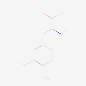
Researchers comparing first-pass metabolism across structurally unrelated compounds frequently run L-DOPA reference material (CAS 59-92-7), whose disposition is dominated by peripheral decarboxylation rather than by conjugation, alongside methylene blue (CAS 61-73-4), which is handled by reduction and demethylation and so occupies a third metabolic category entirely.
References
- Gradolatto A, Basly JP, Berges R, Teyssier C, Chagnon MC, Siess MH, Canivenc-Lavier MC. Pharmacokinetics and metabolism of apigenin in female and male rats after a single oral administration. Drug Metabolism and Disposition. 2005;33(1):49–54. doi:10.1124/dmd.104.000893 · PMID: 15466493
- Gradolatto A, Canivenc-Lavier MC, Basly JP, Siess MH, Teyssier C. Metabolism of apigenin by rat liver phase I and phase II enzymes and by isolated perfused rat liver. Drug Metabolism and Disposition. 2004;32(1):58–65. doi:10.1124/dmd.32.1.58 · PMID: 14709621
- Borges G, Fong RY, Ensunsa JL, Kimball J, Medici V, Ottaviani JI, Crozier A. Absorption, distribution, metabolism and excretion of apigenin and its glycosides in healthy male adults. Free Radical Biology and Medicine. 2022;185:90–96. doi:10.1016/j.freeradbiomed.2022.04.007 · PMID: 35452808
- Meyer H, Bolarinwa A, Wolfram G, Linseisen J. Bioavailability of apigenin from apiin-rich parsley in humans. Annals of Nutrition and Metabolism. 2006;50(3):167–172. doi:10.1159/000090736 · PMID: 16407641
- Nielsen SE, Young JF, Daneshvar B, Lauridsen ST, Knuthsen P, Sandström B, Dragsted LO. Effect of parsley (Petroselinum crispum) intake on urinary apigenin excretion, blood antioxidant enzymes and biomarkers for oxidative stress in human subjects. British Journal of Nutrition. 1999;81(6):447–455. doi:10.1017/s000711459900080x · PMID: 10615220
- Hanske L, Loh G, Sczesny S, Blaut M, Braune A. The bioavailability of apigenin-7-glucoside is influenced by human intestinal microbiota in rats. Journal of Nutrition. 2009;139(6):1095–1102. doi:10.3945/jn.108.102814 · PMID: 19403720
- Chen J, Lin H, Hu M. Metabolism of flavonoids via enteric recycling: role of intestinal disposition. Journal of Pharmacology and Experimental Therapeutics. 2003;304(3):1228–1235. doi:10.1124/jpet.102.046409 · PMID: 12604700
- Hanioka N, Tanaka-Kagawa T, Mori Y, Ikushiro S, Jinno H, Ohkawara S, Isobe T. Regioselective glucuronidation of flavones at C5, C7, and C4′ positions in human liver and intestinal microsomes: comparison among apigenin, acacetin, and genkwanin. Biological and Pharmaceutical Bulletin. 2022;45(8):1116–1123. doi:10.1248/bpb.b22-00160 · PMID: 35908893
- El Daibani AA, Xi Y, Luo L, Mei X, Zhou C, Yasuda S, Liu MC. Sulfation of hesperetin, naringenin and apigenin by the human cytosolic sulfotransferases: a comprehensive analysis. Natural Product Research. 2020;34(6):797–803. doi:10.1080/14786419.2018.1503264 · PMID: 30398375
- Nielsen SE, Dragsted LO. Column-switching high-performance liquid chromatographic assay for determination of apigenin and acacetin in human urine with ultraviolet absorbance detection. Journal of Chromatography B: Biomedical Sciences and Applications. 1998;713(2):379–386. doi:10.1016/s0378-4347(98)00187-x · PMID: 9746253
Research use only. The compounds discussed are supplied as reference standards for laboratory research. They are not medicines, not dietary supplements, and are not for human or animal consumption. Nothing in this article is medical advice or a recommendation for use in humans. Any quantities mentioned are those reported in the published studies cited above and appear solely to describe that research accurately.