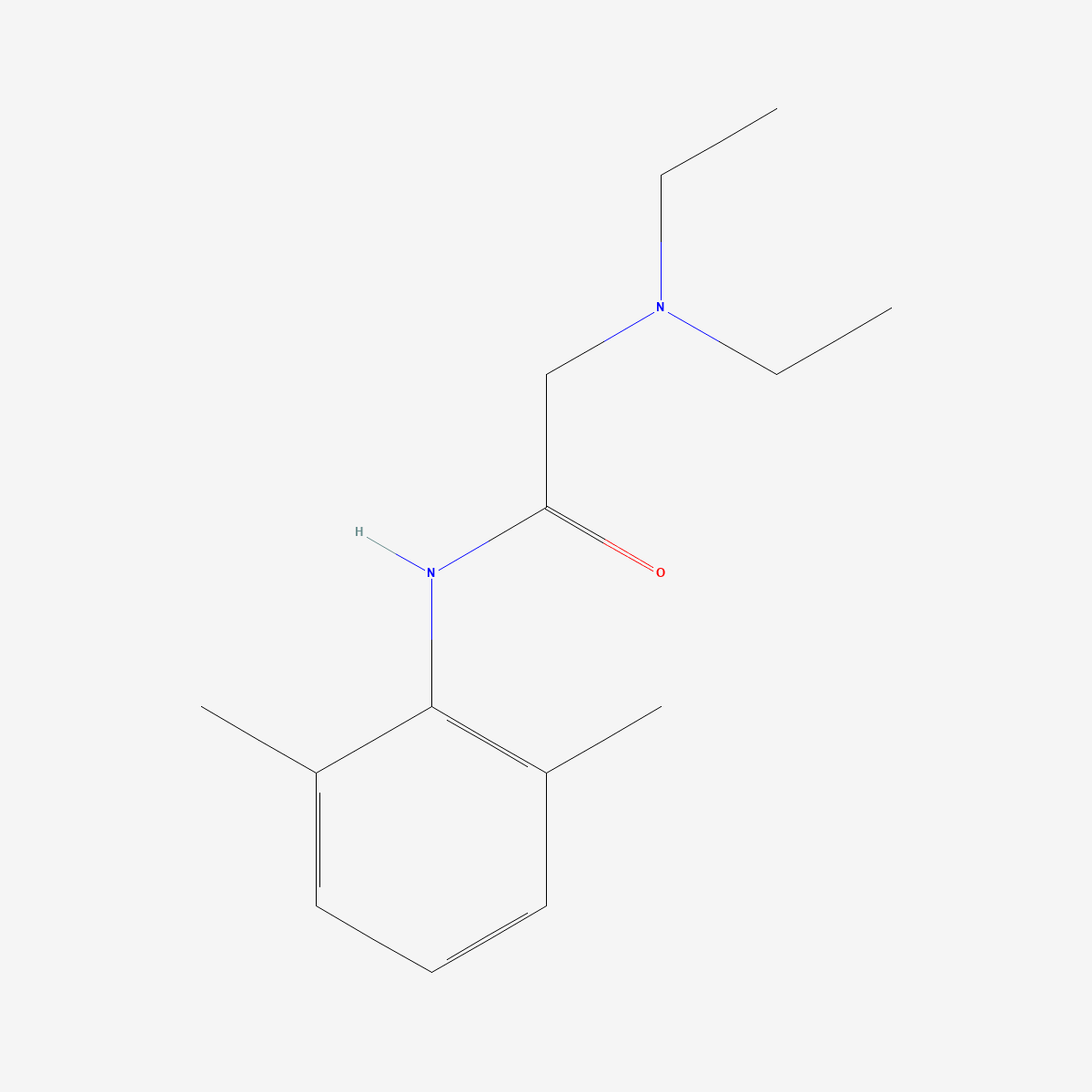
Lidocaine is not given by mouth, and the reason is quantitative rather than traditional: in six healthy subjects given the same dose by three routes, oral systemic availability was 31% measured in whole blood and 34% in plasma, against 63% and 71% by the rectal route. Almost two-thirds of an oral dose is destroyed before it reaches the systemic circulation. That property propagates through the whole pharmacokinetic profile — it is why clearance tracks liver blood flow rather than enzyme capacity, why cardiac failure changes the half-life by a factor of seven, and why the metabolite formed in that first pass became a clinical test of liver function in its own right.
Key points
- Compound: lidocaine (lignocaine), CAS 137-58-6, C14H22N2O, MW 234.34 — an aminoamide local anaesthetic, cleared almost entirely by hepatic metabolism in adults.[1]
- First pass: mean oral systemic availability 31% (whole blood) and 34% (plasma) against 63% and 71% rectally in the same six subjects; a repeat six months later gave 27% orally and 67% rectally.[2]
- Enzyme induction halves it again: oral bioavailability was 0.37 ± 0.09 in normal volunteers and 0.15 ± 0.06 in patients on chronic antiepileptic therapy (P < 0.001), while intravenous clearance did not differ significantly.[3]
- Flow-limited clearance: blood clearance was 11.8 ± 2.6 mL/min/kg in post-infarct patients without heart failure and 7.2 ± 1.9 in those with heart failure (P < 0.002).[5] Half-life was 1.4 hours in healthy subjects, 4.3 hours after infarction without failure, and 10.2 hours with cardiac failure.[4]
- Two active metabolites: monoethylglycinexylidide from N-deethylation, and glycinexylidide, whose elimination depends on renal function and which can accumulate to produce central nervous system toxicity in renal disease even though handling of the parent is unaltered.[6]
- Status: supplied as a reference standard for laboratory research. Not a medicine and not for human or animal consumption.
The first-pass number and how it was obtained
De Boer and colleagues designed the study that settles the question. Six healthy subjects received 200 mg intravenously, 300 mg orally and 300 mg rectally in a balanced crossover, with plasma and whole-blood concentrations measured by capillary gas chromatography. Mean rectal systemic availability exceeded oral: 63% against 31% in whole blood, and 71% against 34% in plasma. Elimination half-lives were about the same intravenously and orally, and slightly longer after rectal administration. The oral and rectal arms were repeated in the same panel about six months later, giving 67% rectally against 27% orally on plasma concentrations, with small intraindividual variability. The authors derived the fraction of a rectal dose bypassing the liver as slightly more than half, assuming complete absorption.[2]
The rectal comparison is what makes the result interpretable. A low oral availability could in principle reflect poor absorption, gut-wall metabolism or hepatic extraction. Recovering most of the dose by a route that partially drains into systemic rather than portal circulation localises the loss to the liver.
Perucca and Richens then showed how sensitive that loss is to enzyme activity. They studied six normal volunteers and six patients on chronic antiepileptic therapy after single oral and intravenous doses. Serum clearance was slightly but not significantly higher in the patients (0.85 ± 0.09 against 0.77 ± 0.07 L/min), yet oral bioavailability differed more than twofold: 0.37 ± 0.09 in the volunteers against 0.15 ± 0.06 in the patients (P < 0.001). The authors attributed the difference to stimulation of hepatic first-pass metabolism by the antiepileptic drugs.[3]
That dissociation — large change in oral bioavailability, negligible change in systemic clearance — is the classic signature of a high-extraction compound. Systemic clearance is already limited by how fast blood delivers drug to the liver, so more enzyme cannot increase it much; but the fraction surviving a single pass through the liver is exquisitely sensitive to enzyme activity.
Clearance limited by delivery, not by enzyme capacity
Oellerich and Armstrong state the principle plainly in their review of the lidocaine-based liver function test: because of the relatively high extraction ratio of lidocaine, the test depends not only on hepatic metabolic capacity but also on hepatic blood flow.[9] The consequences were measured long before the mechanism was framed that way.
Prescott and colleagues infused lignocaine at 1.4 mg/min for 36 to 46 hours in 12 patients with myocardial infarction and one with cardiac failure. In six patients without cardiac failure, plasma concentrations rose progressively during the infusion and the mean half-life was 4.3 hours, against 1.4 hours in healthy subjects. In seven patients with cardiac failure, concentrations were significantly higher and the half-life was prolonged to 10.2 hours. In one patient who developed cardiogenic shock, concentrations rose rapidly to toxic levels and did not fall when the infusion stopped.[4]
Bax and colleagues separated the flow and capacity contributions by measuring lignocaine clearance alongside indocyanine green, a dye whose disposition is used as a marker of hepatic perfusion, in 17 post-infarct patients. Mean lignocaine clearance was 11.8 ± 2.6 mL/min/kg in nine patients without heart failure and 7.2 ± 1.9 in nine with heart failure (P < 0.002). Clearances of the two compounds were not correlated directly, but lignocaine clearance was related to the reciprocal of indocyanine green half-life (rs = 0.67, P < 0.01). Multiple linear regression incorporating indocyanine green half-life and the presence or absence of heart failure accounted for 72% of the variance between observed and predicted lignocaine clearances, but the dye half-life contributed only 17%, which the authors read as showing that a perfusion marker alone is of limited value for predicting lidocaine requirements.[5]
That last finding is the honest version of the flow-limited story. Perfusion is demonstrably part of the explanation but not the whole of it; heart failure alters lidocaine kinetics more than it alters indocyanine green kinetics, so something beyond bulk delivery changes as well.
Kinetics that change during the infusion
Lidocaine is one of the compounds for which single-dose data do not predict steady-state behaviour. Bax and colleagues observed a 25% rise in concentrations between 8–12 hours and 24–28 hours of infusion, and a mean post-infusion terminal half-life of 7.2 ± 2.1 hours in four patients whose infusions ran 30 hours or longer; they concluded that kinetics during and after prolonged infusion were not predicted by bolus data.[5] Prescott and colleagues saw the same progressive rise.[4]
Part of the explanation is protein binding. Routledge and colleagues followed eight patients through a constant 2 mg/min infusion after myocardial infarction, measuring binding, total and free lidocaine, and α1-acid glycoprotein at 12, 24, 36 and 48 hours. By 48 hours total plasma lidocaine, α1-acid glycoprotein and the extent of binding had all risen, and because binding increased in parallel, free concentrations did not change significantly. Correlation between the acute-phase protein and the binding ratio was r = 0.87, and with total plasma concentration r = 0.81.[7]
The implication for measurement is direct: after myocardial infarction, a rising total lidocaine concentration may represent an acute-phase rise in the binding protein rather than accumulation of pharmacologically available drug. Total and free concentration diverge precisely in the population being monitored. A systematic review of published population models lists α1-acid glycoprotein alongside heart failure status, infusion duration and body weight among the significant covariates, and notes that substantial unexplained variability persists even after adjusting for all of them.[11]
Two metabolites and two different problems
Sequential oxidative N-dealkylation gives monoethylglycinexylidide as the major initial metabolite in humans, with glycinexylidide following.[9] Wang and colleagues established which enzymes are responsible, using human liver microsomes and recombinant isoforms. Both CYP1A2 and CYP3A4 catalyse formation of the first metabolite and of 3-hydroxylidocaine. At 5 µM lidocaine — a therapeutically relevant concentration — N-deethylation was inhibited about 60% by furafylline and more than 75% by anti-CYP1A1/2 antibodies, indicating CYP1A2 as the dominant isoform. At 800 µM, troleandomycin inhibited the reaction by about 50%, indicating a greater CYP3A4 contribution. 3-Hydroxylidocaine formation was catalysed almost exclusively by CYP1A2, and CYP2C19, CYP2D6 and CYP2E1 played no significant role.[8]
Isohanni and colleagues tested the assignment in humans. In a double-blind, randomised, three-way crossover, eight healthy volunteers took fluvoxamine 100 mg daily with placebo, fluvoxamine with erythromycin 1500 mg daily, or placebos, for five days, then received oral lidocaine 1 mg/kg on day 6. With fluvoxamine, lidocaine area under the curve was 305% of control (P < 0.001) and maximum concentration 220% (P < 0.05). With the combination, area under the curve was 360% and maximum concentration 250%. Fluvoxamine alone did not significantly change the half-life, but the combination prolonged it from 2.4 to 3.8 hours (P < 0.01). Peak monoethylglycinexylidide concentrations fell by about 50% with fluvoxamine alone and 30% with the combination.[10]
A threefold rise in oral exposure from inhibiting a single enzyme is the first-pass effect measured from the other direction. The second metabolite creates a different problem. Waller’s account of dosing in disease states sets it out: patients with renal disease show no significant alteration in handling of the parent compound, but because glycinexylidide elimination depends on renal function, the metabolite may accumulate and produce central nervous system toxicity.[6] An assay measuring only lidocaine will therefore miss the species responsible for toxicity in exactly the population where it is most likely.
Contemporary modelling handles parent and metabolites jointly. Foong and colleagues studied 34 donor nephrectomy and 64 cholecystectomy patients given intraoperative intravenous lidocaine, analysing 1,520 concentration–time points by non-linear mixed-effects modelling. Lidocaine was best described by a three-compartment model and both metabolites by two-compartment models, with all parameters scaled allometrically on total body mass and fat-free mass. Typical lidocaine clearance was 45.9 L/h with a central volume of 25.2 L — and clearance decreased by 60% postoperatively.[12] That postoperative fall is flow-limited behaviour reappearing in a modern dataset: surgery changes hepatic perfusion, and the clearance of a high-extraction compound follows.
The metabolite as an instrument, and the limits of that instrument
The formation of monoethylglycinexylidide after a test dose of lidocaine became a dynamic liver function test. Oellerich and Armstrong reviewed its application in transplantation and critical care, noting that patients with a 15- or 30-minute test value below 10 µg/L have a particularly poor one-year survival rate, and that an initially rapid decrease in critically ill patients is associated with increased risk of multiple organ dysfunction syndrome. They also flag a measurement caveat: high-performance liquid chromatography and gas–liquid chromatography are specific for the metabolite, whereas the fluorescence polarisation immunoassay cross-reacts with 3-hydroxy-monoethylglycinexylidide — not a problem in humans, but a real one in species such as the rat that produce significant amounts of it.[9]
Wang and colleagues add a second limit, and it is the more important one for experimental design. Because N-deethylation at therapeutic concentrations is predominantly a CYP1A2 reaction, they concluded that the test is not a suitable marker of hepatic CYP3A4 activity in vivo.[8] A detector widely treated as reporting on one enzyme is in fact reporting mainly on another, and which isoform dominates changes across the concentration range.
None of this bears on how lidocaine acts at its molecular target, which is covered separately in our discussion of state-dependent block of the voltage-gated sodium channel. The point here is narrower: the disposition is interesting enough to have been repurposed as a measuring device, and the device inherits every one of its complications.
Frequently asked questions
Why is lidocaine not given by mouth?
Because most of an oral dose does not survive the first pass through the liver. In a balanced crossover in six healthy subjects, mean oral systemic availability was 31% in whole blood and 34% in plasma, against 63% and 71% by the rectal route, which partially bypasses portal drainage.[2] In patients on enzyme-inducing antiepileptic therapy, oral bioavailability fell to 0.15 ± 0.06 while systemic clearance was essentially unchanged.[3]
What is the half-life of lidocaine?
It depends on the circulation. Roughly 1.4 hours in healthy subjects, 4.3 hours in patients after myocardial infarction without cardiac failure, and 10.2 hours in patients with cardiac failure.[4] After prolonged infusion the post-infusion terminal half-life was 7.2 ± 2.1 hours in patients whose infusions had run 30 hours or more.[5] A single quoted half-life for this compound is close to meaningless without the clinical state attached.
Why do the metabolites matter?
Both are pharmacologically relevant and they behave differently. Monoethylglycinexylidide is formed rapidly by CYP1A2 at therapeutic concentrations and by CYP3A4 at high concentrations,[8] and its formation rate is used as a liver function test.[9] Glycinexylidide depends on renal function for elimination and can accumulate in renal disease to produce central nervous system toxicity, even though handling of the parent compound is not significantly altered.[6] Modern population models fit parent and both metabolites jointly for this reason.[12]
What should a lidocaine reference standard provide for pharmacokinetic work?
A stated assay with the method behind it, and explicit identification of salt form, because free base and hydrochloride differ in molar content per unit mass and the difference propagates into every calculated concentration. Our lidocaine reference standard (CAS 137-58-6) is supplied with identity and purity documentation available on request.
Related compounds in our catalogue
Comparative work on local anaesthetic disposition pairs this compound with procaine hydrochloride reference material (CAS 51-05-8), an ester agent hydrolysed by plasma cholinesterase rather than cleared hepatically,[1] and with benzocaine reference standard (CAS 94-09-7), which lacks the ionisable amine and so differs in both distribution and elimination.
References
- Tucker GT, Mather LE. Clinical pharmacokinetics of local anaesthetics. Clinical Pharmacokinetics. 1979;4(4):241–278. doi:10.2165/00003088-197904040-00001 · PMID: 385208
- de Boer AG, Breimer DD, Mattie H, Pronk J, Gubbens-Stibbe JM. Rectal bioavailability of lidocaine in man: partial avoidance of “first-pass” metabolism. Clinical Pharmacology and Therapeutics. 1979;26(6):701–709. doi:10.1002/cpt1979266701 · PMID: 498711
- Perucca E, Richens A. Reduction of oral bioavailability of lignocaine by induction of first pass metabolism in epileptic patients. British Journal of Clinical Pharmacology. 1979;8(1):21–31. doi:10.1111/j.1365-2125.1979.tb05904.x · PMID: 552293
- Prescott LF, Adjepon-Yamoah KK, Talbot RG. Impaired lignocaine metabolism in patients with myocardial infarction and cardiac failure. British Medical Journal. 1976;1(6015):939–941. doi:10.1136/bmj.1.6015.939 · PMID: 1268491
- Bax ND, Tucker GT, Woods HF. Lignocaine and indocyanine green kinetics in patients following myocardial infarction. British Journal of Clinical Pharmacology. 1980;10(4):353–361. doi:10.1111/j.1365-2125.1980.tb01771.x · PMID: 7448106
- Waller ES. Pharmacokinetic principles of lidocaine dosing in relation to disease state. Journal of Clinical Pharmacology. 1981;21(4):181–194. doi:10.1002/j.1552-4604.1981.tb05698.x · PMID: 7240439
- Routledge PA, Shand DG, Barchowsky A, Wagner G, Stargel WW. Relationship between alpha 1-acid glycoprotein and lidocaine disposition in myocardial infarction. Clinical Pharmacology and Therapeutics. 1981;30(2):154–157. doi:10.1038/clpt.1981.141 · PMID: 7249498
- Wang JS, Backman JT, Taavitsainen P, Neuvonen PJ, Kivistö KT. Involvement of CYP1A2 and CYP3A4 in lidocaine N-deethylation and 3-hydroxylation in humans. Drug Metabolism and Disposition. 2000;28(8):959–965. PMID: 10901707
- Oellerich M, Armstrong VW. The MEGX test: a tool for the real-time assessment of hepatic function. Therapeutic Drug Monitoring. 2001;23(2):81–92. doi:10.1097/00007691-200104000-00001 · PMID: 11294522
- Isohanni MH, Neuvonen PJ, Olkkola KT. Effect of fluvoxamine and erythromycin on the pharmacokinetics of oral lidocaine. Basic & Clinical Pharmacology & Toxicology. 2006;99(2):168–172. doi:10.1111/j.1742-7843.2006.pto_482.x · PMID: 16918719
- Foong KW, Chaw SH, Lo YL, Loh PS. Population pharmacokinetics of intravenous lidocaine in adults: a systematic review. Clinical Pharmacokinetics. 2024;63(5):623–643. doi:10.1007/s40262-024-01373-4 · PMID: 38703307
- Foong KW, Loh PS, Chaw SH, Lo YL. Population pharmacokinetics of IV lidocaine and its metabolites in adult surgical patients. Pharmaceutical Research. 2025;42(3):451–473. doi:10.1007/s11095-025-03835-1 · PMID: 40021547
Research use only. The compounds discussed are supplied as reference standards for laboratory research. They are not medicines, not dietary supplements, and are not for human or animal consumption. Nothing in this article is medical advice or a recommendation for use in humans. Any quantities mentioned are those reported in the published studies cited above and appear solely to describe that research accurately.