Ask what concentration of lidocaine blocks half of a population of sodium channels and the honest answer is a question in return: at what membrane potential, and after what stimulus history. In voltage-clamped cardiac Purkinje fibres the half-blocking concentration moved from above 300 µM at a holding potential where inactivation was fully removed to roughly 10 µM where inactivation was nearly complete — a thirty-fold shift in the same preparation, with the same drug. Lidocaine does not have an affinity for the sodium channel. It has an affinity for each of the channel’s conformations, and the tissue decides which one the drug meets.
Key points
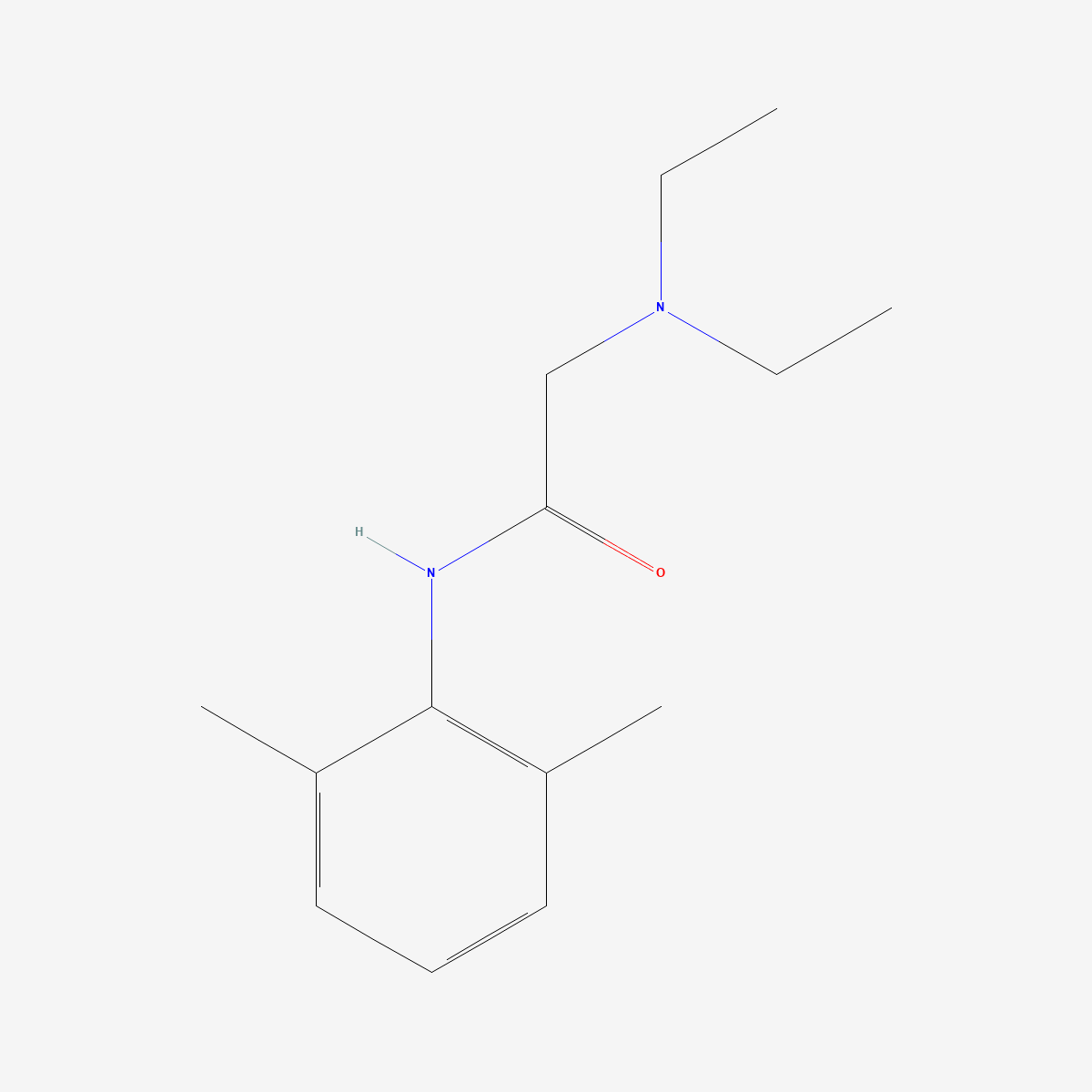
- Structure: lidocaine is 2-(diethylamino)-N-(2,6-dimethylphenyl)acetamide (CAS 137-58-6, C14H22N2O, MW 234.34) — an amide-linked local anaesthetic with an ionisable tertiary amine, so both neutral and protonated species are present near physiological pH.
- Target: the local anaesthetic receptor in the inner pore of voltage-gated sodium channels, reached by two distinct routes that depend on the drug’s charge state.[1]
- Defining property: state-dependent, use-dependent block. Affinity for inactivated channels is far higher than for resting channels, so block accumulates with depolarisation and with stimulus frequency.[2]
- Molecular anchor: two aromatic residues in transmembrane segment IVS6 — F1764 and Y1771 in the rat type IIA numbering, Phe1759 in NaV1.5 — dominate high-affinity state-dependent binding.[3][4]
- Non-channel actions: interference with Gαq-mediated receptor signalling has been demonstrated at an intracellular site, distinct from pore block.[7]
- Status: supplied as a reference standard for laboratory research. Not a medicine, not a supplement, and not for human or animal consumption.
One receptor, two ways in
The framework still used to interpret local anaesthetic data was set out in 1977 on the node of Ranvier. Comparing neutral, ionisable-amine and permanently charged quaternary compounds, Hille found that conventional 50 ms prepulse protocols showed a large negative shift of the steady-state inactivation curve with neutral benzocaine and with ionisable amines such as lidocaine and tetracaine, but no shift with the quaternary compound QX-572 — unless the protocol was changed to repetitive prepulse–testpulse stimulation, at which point the quaternary shift appeared as well. One-minute hyperpolarisations restored two to four times as many channels to the conducting pool as 50 ms hyperpolarisations in fibres treated with lidocaine or tetracaine, but did not relieve block by internally applied quaternary drugs.[1]
The interpretation was a single receptor reached by two pathways: lipid-soluble forms arriving through a hydrophobic region of the membrane, charged and less lipid-soluble forms through a hydrophilic route at the inner channel mouth that is open only when the channel’s gates are open. Any drug occupying the site then raises the probability that the inactivation gate closes — which is equivalent to shifting the voltage dependence of inactivation in the negative direction. Everything that followed in this field is an elaboration or a test of that sentence.
A dissociation constant that moves with voltage
The quantitative demonstration came from voltage-clamped rabbit Purkinje fibres studied across concentrations from 1 mM down to the low micromolar range. Dose–response analysis indicated one-to-one binding with a voltage-dependent dissociation constant: the half-blocking concentration ran from more than 300 µM at a negative holding potential where inactivation was completely removed to approximately 10 µM at a depolarised holding potential where inactivation was nearly complete.[2]
Recovery of the sodium current between pulses separated into two exponential components — a normally recovering component with a time constant under 0.2 s, and a lidocaine-induced slowly recovering fraction with a time constant of roughly 1–2 s at pH 7.0. Raising the concentration made the slow fraction larger without changing its time course, and after a long depolarisation that fraction reached one half at about 10 µM, exactly as expected if it corresponded to drug-bound inactivated channels. At 20 µM and below, the slow fraction grew with the duration of the preceding depolarisation along the same time course whether the depolarisation was strong or weak — that is, with or without significant channel opening. The authors drew the conclusion that matters for interpreting any use-dependence experiment: at therapeutic concentrations, use dependence reflects block of inactivated channels rather than block of open channels.[2]
Locating the site: mutagenesis before structures
Site-directed mutagenesis identified the anchor points nearly two decades before any sodium channel structure existed. In transmembrane segment IVS6 of type IIA channel α subunits, the mutation F1764A reduced the affinity of inactivated channels for lidocaine 24.5-fold and for phenytoin 8.3-fold, while Y1771A had smaller effects. For quinidine and flecainide, which prefer open channels, the same mutations reduced open-state affinity only two- to three-fold. The conclusion was a common receptor site engaged in an overlapping but non-identical way by chemically diverse drugs — local anaesthetics, class I antiarrhythmics and anticonvulsants alike.[3]
Work on the cardiac channel then separated the pharmacology into two measurable components. Replacing Phe1759 of NaV1.5 with a non-aromatic residue split the actions of lidocaine and benzocaine into a voltage-independent, low-affinity block of closed channels, and a high-affinity, voltage-dependent block of open and inactivated channels linked to stabilisation of the domain III and domain IV voltage sensors. The authors named these lipophilic block and voltage-sensor inhibition, and argued that the difference in affinity between the two binding conformations is what controls use dependence — described in that paper as the hallmark of successful antiarrhythmic drugs.[4]
The structures caught up with Hille
Rosetta modelling and molecular dynamics simulations of antiarrhythmic and local anaesthetic interactions with human NaV1.5 revealed several drug binding sites within the pore lumen, capable of accommodating up to two drug molecules simultaneously, and identified two access routes: a hydrophilic pathway through the intracellular gate and a hydrophobic pathway through a fenestration between domains III and IV.[5] That is the 1977 hypothesis, recovered from atomic-scale simulation rather than from gating kinetics.
Experimental structures of NaV1.5 at 3.2–3.5 Å resolution followed, captured with voltage sensors partially activated and the fast-inactivation gate partially closed, and showed the antiarrhythmic flecainide specifically occupying the central cavity of the pore.[6] Note what this does and does not settle: it fixes the geometry of the site, but a single static structure cannot supply the state-dependent affinity ratios that the electrophysiology measured. The kinetic work is not superseded by the structural work; the two answer different questions.
Actions that are not pore block
Local anaesthetics are described as sodium channel blockers, but effects at concentrations and timescales that pore block does not explain have been reported for decades. One study characterised the responsible site directly. In Xenopus oocytes, ropivacaine inhibited lysophosphatidate receptor signalling stereoselectively and non-competitively — the signature of an interaction with a protein rather than with membrane lipid. Antisense work then identified Gαq among the primary subunits mediating lysophosphatidate, m1 muscarinic and trypsin receptor signalling, all of which were inhibited to a similar degree by intracellularly injected QX314, a permanently charged lidocaine analogue. The angiotensin1A receptor, previously shown to be unaffected by local anaesthetics, signalled through Gαo and Gα14 rather than Gαq — which is the negative control that makes the argument work.[7]
How much of the clinical picture this mechanism carries is genuinely unresolved. A Cochrane review of 68 randomised trials in 4525 participants found the evidence for continuous intravenous perioperative lidocaine infusion mixed: uncertain effect on early postoperative pain at 1–4 hours (SMD −0.50, 95% CI −0.72 to −0.28; 29 studies, 1656 participants; very low-quality evidence), with a clinically relevant reduction ruled out at 24 hours (SMD −0.14, 95% CI −0.25 to −0.04; moderate quality) and at 48 hours. Infusion regimens across the included studies varied from 1 to 5 mg/kg/h and terminated anywhere from the end of surgery to several days later.[8] That heterogeneity is itself informative: a systemic mechanism with a defined molecular target should be easier to pin down than this.
Practical notes for laboratory handling
Lidocaine free base and lidocaine hydrochloride behave very differently in aqueous work, and which one a protocol assumes is worth checking before a solution is made. The free base is the lipophilic species relevant to Hille’s hydrophobic access route; the salt is the water-soluble form. Because block depends on the ratio of neutral to protonated drug, buffer pH is an experimental variable in its own right rather than a background detail, and the pH of the bath should be reported with any affinity estimate. The amide linkage is markedly more stable to hydrolysis than the ester linkage of procaine-type anaesthetics, which is why the two classes differ in shelf behaviour as much as in metabolic fate.
Frequently asked questions
What does use-dependent block actually mean?
It means the fraction of blocked channels grows with repeated depolarisation rather than being set solely by concentration. The mechanism is that the drug binds inactivated channels with far higher affinity than resting ones and unbinds slowly — in Purkinje fibres the lidocaine-induced recovery component had a time constant of about 1–2 s at pH 7.0, against under 0.2 s for normal repriming.[2] Rapidly firing tissue therefore accumulates block that quiescent tissue does not.
Is lidocaine selective for cardiac sodium channels?
No. The 1983 study explicitly noted that, unlike tetrodotoxin, lidocaine shows similar interactions with sodium channels of heart, nerve and skeletal muscle.[2] Apparent tissue selectivity in practice comes from differences in firing pattern and resting potential, not from differences in the binding site.
Why do local anaesthetics and class I antiarrhythmics share a binding site?
Because they are the same pharmacology applied to different tissues. Mutating F1764 and Y1771 reduced state-dependent block by lidocaine, phenytoin, quinidine and flecainide together, though to different extents, indicating an overlapping but non-identical interaction with one receptor site in the pore.[3]
What purity is appropriate for lidocaine used as an analytical reference?
For quantitative electrophysiology and for chromatographic calibration, material characterised at ≥99% with a stated assay method is the working minimum, and the salt form must be stated explicitly since it changes both the molar mass used in calculations and the aqueous behaviour of the stock. Our lidocaine reference standard (CAS 137-58-6) is supplied with identity documentation available on request.
Related compounds in our catalogue
Researchers separating the charge-dependent and charge-independent components of local anaesthetic action commonly pair lidocaine with benzocaine reference material (CAS 94-09-7), the neutral compound used alongside lidocaine in the NaV1.5 conformational work discussed above, and with procaine hydrochloride (CAS 51-05-8), an ester-linked anaesthetic that differs from lidocaine in hydrolytic stability rather than in target.
References
- Hille B. Local anesthetics: hydrophilic and hydrophobic pathways for the drug-receptor reaction. Journal of General Physiology. 1977;69(4):497–515. doi:10.1085/jgp.69.4.497 · PMID: 300786
- Bean BP, Cohen CJ, Tsien RW. Lidocaine block of cardiac sodium channels. Journal of General Physiology. 1983;81(5):613–642. doi:10.1085/jgp.81.5.613 · PMID: 6306139
- Ragsdale DS, McPhee JC, Scheuer T, Catterall WA. Common molecular determinants of local anesthetic, antiarrhythmic, and anticonvulsant block of voltage-gated Na+ channels. Proceedings of the National Academy of Sciences of the USA. 1996;93(17):9270–9275. doi:10.1073/pnas.93.17.9270 · PMID: 8799190
- Hanck DA, Nikitina E, McNulty MM, Fozzard HA, Lipkind GM, Sheets MF. Using lidocaine and benzocaine to link sodium channel molecular conformations to state-dependent antiarrhythmic drug affinity. Circulation Research. 2009;105(5):492–499. doi:10.1161/CIRCRESAHA.109.198572 · PMID: 19661462
- Nguyen PT, DeMarco KR, Vorobyov I, Clancy CE, Yarov-Yarovoy V. Structural basis for antiarrhythmic drug interactions with the human cardiac sodium channel. Proceedings of the National Academy of Sciences of the USA. 2019;116(8):2945–2954. doi:10.1073/pnas.1817446116 · PMID: 30728299
- Jiang D, Shi H, Tonggu L, Gamal El-Din TM, Lenaeus MJ, Zhao Y, Yoshioka C, Zheng N, Catterall WA. Structure of the cardiac sodium channel. Cell. 2020;180(1):122–134.e10. doi:10.1016/j.cell.2019.11.041 · PMID: 31866066
- Hollmann MW, Wieczorek KS, Berger A, Durieux ME. Local anesthetic inhibition of G protein-coupled receptor signaling by interference with Galpha(q) protein function. Molecular Pharmacology. 2001;59(2):294–301. doi:10.1124/mol.59.2.294 · PMID: 11160866
- Weibel S, Jelting Y, Pace NL, Helf A, Eberhart LH, Hahnenkamp K, Hollmann MW, Poepping DM, Schnabel A, Kranke P. Continuous intravenous perioperative lidocaine infusion for postoperative pain and recovery in adults. Cochrane Database of Systematic Reviews. 2018;6(6):CD009642. doi:10.1002/14651858.CD009642.pub3 · PMID: 29864216
Research use only. The compounds discussed are supplied as reference standards for laboratory research. They are not medicines, not dietary supplements, and are not for human or animal consumption. Nothing in this article is medical advice or a recommendation for use in humans. Any quantities mentioned are those reported in the published studies cited above and appear solely to describe that research accurately.