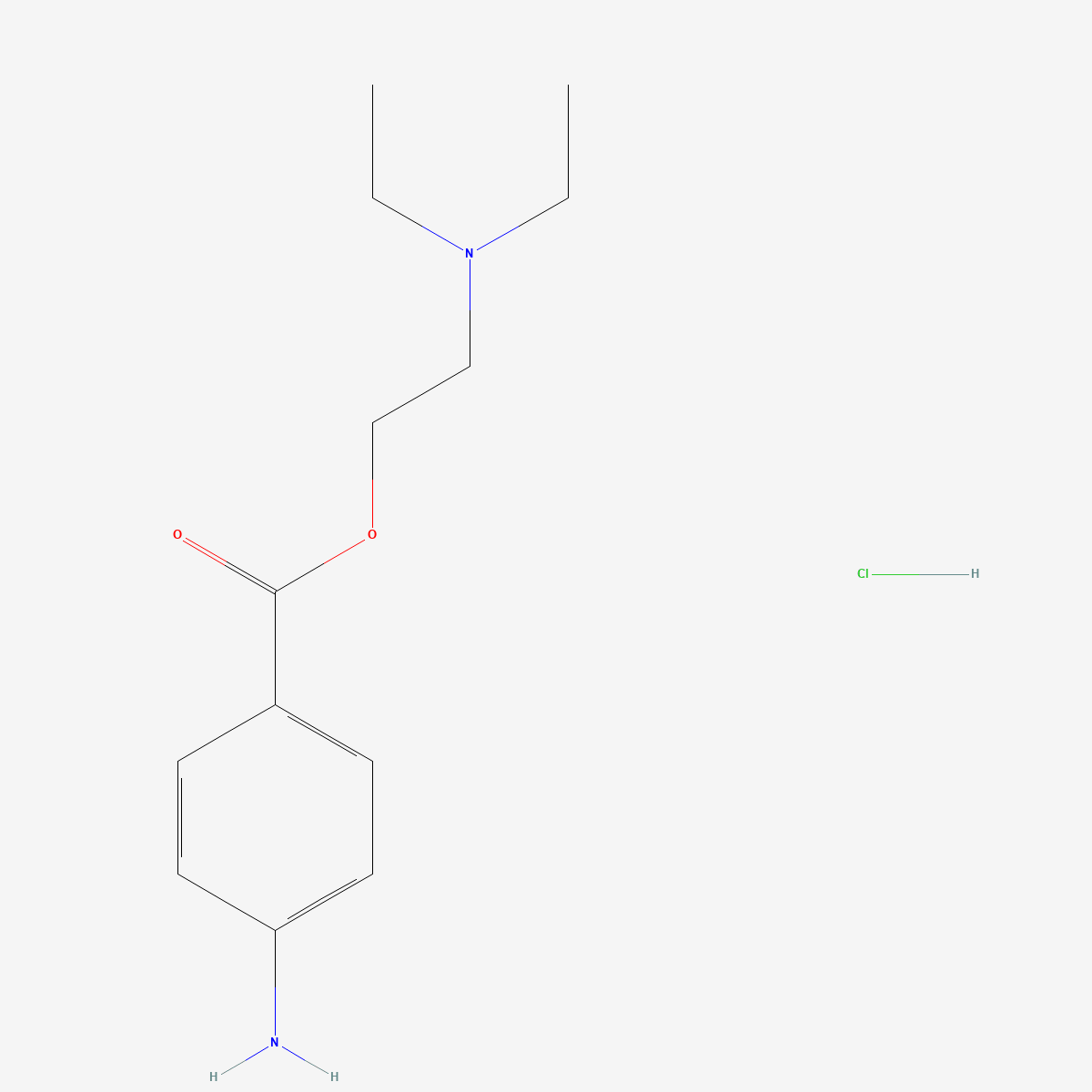
Procaine is the molecule that ended the cocaine era, and it did so by solving two problems at once with two functional groups. A tertiary amine on a short side chain gives it water solubility and a positive charge at physiological pH — which dictates how it reaches the sodium channel. An ester link to a para-aminobenzoate ring gives it a scheduled exit: esterases in plasma and tissue cleave it, and what remains is para-aminobenzoic acid. Nearly everything distinctive about procaine, from its use-dependent block to its short duration to its reputation for allergy, traces back to one or the other of those two groups.
Key points
- Structure: 2-(diethylamino)ethyl 4-aminobenzoate hydrochloride (CAS 51-05-8, C13H21ClN2O2, MW 272.77). The free base is C13H20N2O2, MW 236.31 — about 86.6% of the salt mass.
- Access route: as a protonated amine at physiological pH, procaine reaches the sodium channel receptor mainly through the hydrophilic pathway at the inner channel mouth, which is open only while the gates are open.[4]
- Kinetics at the channel: in voltage-clamped frog nodes, 1.0 mM procaine at pH 7.2 gave mean half-times of 3.7 s for onset and 28 s for offset of block, with concentration dependence consistent with a one-to-one drug–receptor reaction.[5]
- Metabolism is the mechanism: ester hydrolysis by carboxylesterases — including in skin, where inhibition by loperamide implicates human hCE2 — removes the molecule and generates para-aminobenzoic acid.[6]
- The allergy assumption is not supported by the controlled data: 340 intracutaneous injections in 177 patients produced no type-1 allergy to procaine, lidocaine or mepivacaine; procaine’s larger wheals reflected vasodilatation.[9]
- A second, unrelated literature: procaine binds CpG-enriched DNA and produced a 40% reduction in 5-methylcytosine content in MCF-7 cells, with growth inhibition and mitotic arrest.[10]
- Status: supplied as a reference standard for laboratory research. Not a medicine and not for human or animal consumption.
The molecule that replaced cocaine
The historical arc is compact and well documented. Coca leaves reached Europe in quantity in 1850, cocaine was isolated from them, and in 1884 Koller performed the first clinical operation under local anaesthesia by applying cocaine to the eye. Use spread rapidly across Europe and America, and so did the deaths — among patients and among addicted medical staff. Local anaesthesia was in a profound crisis until organic chemistry offered replacements, and a series of amino ester anaesthetics followed between 1891 and 1930.[1] Alfred Einhorn is credited with the discovery of procaine.[2]
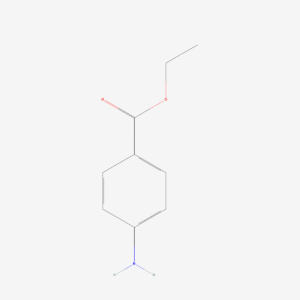
The design that emerged is worth looking at as a design. Take a para-aminobenzoate ester — the same aromatic core as benzocaine, the uncharged member of the family — and replace the plain ethyl ester with a 2-(diethylamino)ethyl group. That single substitution adds a tertiary aliphatic amine, which is substantially protonated at pH 7.4, which in turn makes the molecule water-soluble as a hydrochloride salt and injectable. It also completely changes how the molecule gets to its target.
Why the charge changes the route
Hille’s study of sodium channels at the node of Ranvier established the framework still used today. Neutral, ionisable amine and permanently quaternary local anaesthetics were compared under voltage clamp. Neutral benzocaine and ionisable amines such as lidocaine and tetracaine produced a large negative shift of the steady-state inactivation curve with conventional 50 ms prepulses; permanently charged QX-572 did not, unless the experiment was run with repetitive prepulse–testpulse waveforms. Long hyperpolarisations and raised external calcium relieved block by the ionisable amines but not by internal quaternary drugs. Hille’s interpretation was a single receptor approached by two routes: lipid-soluble forms through a hydrophobic region of the membrane, charged and less lipid-soluble forms through a hydrophilic region — the inner channel mouth — which is open only when the gates of the channel are open.[4] Narahashi and colleagues had approached the same question through pH experiments on tertiary compounds.[3]
Place procaine in that scheme and its behaviour stops being a list of clinical facts and becomes a consequence. A drug that must pass through an open gate to reach its site will bind preferentially to channels that are firing, which is the definition of use-dependent block. It will also be sensitive to extracellular pH, because pH determines what fraction of the dose is in the lipid-permeant neutral form available to cross the membrane in the first place.
Kinetics at the channel, and where procaine does not bind
Wagner and Ulbricht measured procaine block directly in single myelinated nerve fibres from Rana esculenta. Following changes in sodium current during repetitive 1 Hz depolarising pulses after hyperpolarising prepulses, 1.0 mM procaine at pH 7.2 gave mean half-times of 3.7 s for onset and 28 s for offset. Concentrations from 0.03 to 1.0 mM gave equilibrium effects consistent with a one-to-one drug–receptor reaction.[5]
The more instructive half of that experiment was the control. In the presence of 1.4 nM saxitoxin, the onset and offset half-times were essentially unchanged at 3.1 and 27 s, and the partial relief of procaine block obtained by raising the pulse frequency from 1 to 10 Hz was likewise unaffected. The fraction of channels blocked at equilibrium was 0.81 with procaine alone, 0.49 with saxitoxin alone and 0.90 with both together — the arithmetic of two independent receptors, not one.[5] Procaine does not occupy the outer guanidinium toxin site that saxitoxin binds. It reaches its receptor by the route Hille assigned to charged drugs — the inner channel mouth.
The ester bond is half the pharmacology
Procaine’s short duration of action is usually stated as a clinical fact. It is really an enzymology result. Jewell and colleagues compared the capacity of human, minipig and rat skin and liver subcellular fractions to hydrolyse procaine against a panel of carboxylesterase and arylesterase substrates. Rates of procaine hydrolysis by minipig and human skin microsomal and cytosolic fractions were similar, with rat markedly higher. Loperamide inhibited procaine hydrolysis by human skin, implicating human carboxylesterase hCE2. Minipig and human liver and skin esterase activity was inhibited principally by paraoxon and bis-nitrophenyl phosphate, the classical carboxylesterase inhibitors, whereas rat activity was additionally inhibited by phenylmethylsulfonyl fluoride and by the arylesterase inhibitor mercuric chloride — a different enzyme profile altogether. The authors drew the practical conclusion directly: skin has the capacity to hydrolyse procaine following topical application, which possibly limits its pharmacological effect.[6]
Two things follow for experimental design. First, species choice is not neutral: rat skin and liver metabolise esters faster and through a partly different enzyme complement than human tissue, while minipig resembles human closely. Second, the tissue itself is part of the assay. A topical procaine experiment measures delivery and hydrolysis at the same time, and the two cannot be separated by measuring effect alone.
para-Aminobenzoic acid is both the metabolite and the stability problem
The hydrolysis product is para-aminobenzoic acid (PABA), and it appears by two quite different routes: enzymatically in tissue, and non-enzymatically in the bottle. PABA is characterised in the analytical literature as a related substance and degradation product of procaine hydrochloride, and can be measured in the presence of the parent by zero-crossing first-derivative spectrometry at 290 nm, with calibration linear up to 10 mg L−1 and a detection limit of 0.70 mg L−1, without sample pretreatment.[7]
Where sensitivity matters, a validated LC-MS/MS method determines procaine and PABA simultaneously using N-acetylprocainamide as internal standard, on an XTerra MS C18 column with positive-mode electrospray and multiple reaction monitoring at m/z 237 → 100 for procaine, 138 → 120 for PABA and 278 → 205 for the internal standard. Retention times were 4.0, 4.7 and 5.8 min respectively; linearity ran from 10 to 5000 nM for procaine and 100 to 5000 nM for PABA, with intra- and inter-day relative standard deviations below 5%.[8]
The practical message for anyone storing aqueous procaine is that an old solution is not a weaker procaine solution — it is a mixture of procaine and PABA, and the second component is not pharmacologically silent. If your protocol depends on concentration, measure it rather than assuming it.
The allergy reputation, and what a controlled study found
Ester-linked local anaesthetics are routinely avoided on the grounds of high allergenicity, an assumption usually attributed to the PABA metabolite. Weinschenk and colleagues tested it prospectively. In a double-blinded, placebo-controlled design, 177 patients received 340 intracutaneous injections of 1% procaine, 0.5% lidocaine, 1% mepivacaine or saline, two different tests per forearm, monitored for 15 minutes. No type-1 allergy was observed. Mean erythema diameter at 10 minutes was 8.0 ± 6.4 mm for procaine, 7.6 ± 6.3 mm for mepivacaine, 4.4 ± 4.8 mm for lidocaine and 3.7 ± 3.2 mm for saline, with procaine and mepivacaine wheals significantly larger than those of lidocaine and saline. No general signs of hypersensitivity were seen, and wheal diameter and intensity were independent of injection site, order, age, sex and body mass index. The authors attributed the larger wheals to vasodilatation, and concluded that the ester-linked anaesthetic showed no higher type-1 allergenicity than the amides.[9]
One controlled study does not settle a question of this size, and the design measured immediate-type reactivity in a specific setting. But it is the kind of evidence that ought to displace an assumption repeated for decades without measurement, and it illustrates a general point worth carrying into any reading of this literature: a visible skin reaction is not the same entity as an immunological one.
A second literature that has nothing to do with nerves
Methylation-associated silencing of tumour suppressor genes is a recognised feature of human cancer, and because methylation is reversible it has attracted interest as a target. Following the observation that the antiarrhythmic procainamide is a non-nucleoside inhibitor of DNA methyltransferases, Villar-Garea and colleagues tested procaine. In MCF-7 breast cancer cells, procaine produced a 40% reduction in 5-methylcytosine DNA content as measured by high-performance capillary electrophoresis and by total DNA enzyme digestion, demethylated densely hypermethylated CpG islands including the RARβ2 promoter with restoration of gene expression, and showed growth-inhibitory effects with mitotic arrest. The authors also found that procaine binds CpG-enriched DNA, which they proposed as the explanation.[10]
This is a completely separate research use for the same reagent, and it is the reason procaine turns up in epigenetics laboratories that have no interest in sodium channels. It is also a reminder that “the mechanism of procaine” is an ambiguous phrase: the concentrations and timescales in the two literatures do not overlap.
Frequently asked questions
What is the difference between procaine and procaine hydrochloride?
They are the free base and its hydrochloride salt of the same molecule. The base is C13H20N2O2, MW 236.31; the hydrochloride is C13H21ClN2O2, MW 272.77. A weighed portion of the salt therefore delivers about 86.6% of that mass as base. The salt is what is normally supplied and handled, because it is the water-soluble form.
Why does procaine wear off faster than lidocaine?
Because of the bond type, not the potency. Procaine is an ester and is cleaved by carboxylesterases in plasma and tissue, including in skin, which the metabolism literature identifies as a factor that possibly limits its effect after topical application.[6] Lidocaine is an amide and is not a substrate for those enzymes; its clearance depends on hepatic metabolism instead. Substituting an amide for the ester is the single change that separates the two families.
Does procaine really cause more allergic reactions than amide anaesthetics?
The controlled data available do not support the claim. A double-blinded, placebo-controlled study of 340 intracutaneous injections found no type-1 allergy to procaine, lidocaine or mepivacaine, and attributed the larger procaine wheals to vasodilatation rather than hypersensitivity.[9] That is one study in one setting, and it is not clinical advice, but the assumption it tested had never been measured as carefully.
What should a procaine hydrochloride reference standard be characterised for?
Assay with the method stated, explicit salt form, and a figure for free PABA, since that is the expected degradation product and it accumulates in aqueous solution over time.[7] Our procaine hydrochloride reference standard (CAS 51-05-8) ships with identity documentation available on request.
Related compounds in our catalogue
The two compounds that make procaine interpretable in a comparison set are benzocaine (CAS 94-09-7), the same aromatic ester stripped of its basic side chain and therefore uncharged at physiological pH, and lidocaine reference material (CAS 137-58-6), which keeps the basic amine but replaces the ester with an amide and so isolates the metabolic variable.
References
- Ruetsch YA, Böni T, Borgeat A. From cocaine to ropivacaine: the history of local anesthetic drugs. Current Topics in Medicinal Chemistry. 2001;1(3):175–182. doi:10.2174/1568026013395335
- Dunsky JL. Alfred Einhorn: the discoverer of procaine. Journal of the Massachusetts Dental Society. 1997;46(3):25–26. PMID: 9540713
- Narahashi T, Frazier T, Yamada M. The site of action and active form of local anesthetics. I. Theory and pH experiments with tertiary compounds. Journal of Pharmacology and Experimental Therapeutics. 1970;171(1):32–44. PMID: 5410936
- Hille B. Local anesthetics: hydrophilic and hydrophobic pathways for the drug-receptor reaction. Journal of General Physiology. 1977;69(4):497–515. doi:10.1085/jgp.69.4.497
- Wagner HH, Ulbricht W. Saxitoxin and procaine act independently on separate sites of the sodium channel. Pflügers Archiv. 1976;364(1):65–70. doi:10.1007/BF01062913
- Jewell C, Ackermann C, Payne NA, Fate G, Voorman R, Williams FM. Specificity of procaine and ester hydrolysis by human, minipig, and rat skin and liver. Drug Metabolism and Disposition. 2007;35(11):2015–2022. doi:10.1124/dmd.107.015727
- Badea I, Moja D, Vladescu L. Determination of para-aminobenzoic acid, a degradation product of procaine hydrochloride, by zero-crossing first-derivative spectrometry. Analytical and Bioanalytical Chemistry. 2002;374(1):51–53. doi:10.1007/s00216-002-1446-0
- Dhananjeyan MR, Bykowski C, Trendel JA, Sarver JG, Ando H, Erhardt PW. Simultaneous determination of procaine and para-aminobenzoic acid by LC-MS/MS method. Journal of Chromatography B. 2007;847(2):224–230. doi:10.1016/j.jchromb.2006.10.004
- Weinschenk S, Mergenthaler C, Armstrong C, Göllner R, Hollmann MW, Strowitzki T. Local anesthetics, procaine, lidocaine, and mepivacaine show vasodilatation but no type 1 allergy: a double-blind, placebo-controlled study. BioMed Research International. 2017;2017:9804693. doi:10.1155/2017/9804693
- Villar-Garea A, Fraga MF, Espada J, Esteller M. Procaine is a DNA-demethylating agent with growth-inhibitory effects in human cancer cells. Cancer Research. 2003;63(16):4984–4989. PMID: 12941824
Research use only. The compounds discussed are supplied as reference standards for laboratory research. They are not medicines, not dietary supplements, and are not for human or animal consumption. Nothing in this article is medical advice or a recommendation for use in humans. Any quantities mentioned are those reported in the published studies cited above and appear solely to describe that research accurately.